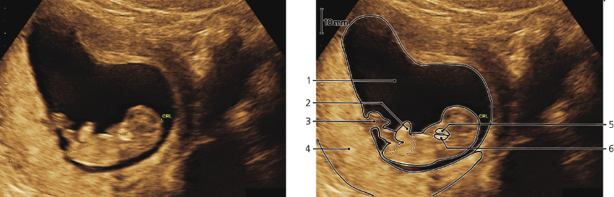
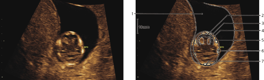
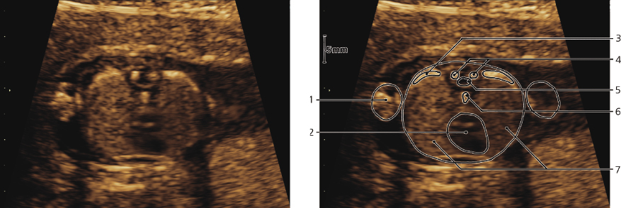
GA: 9w4d, CRL: 23 mm
- Decidua capsularis
- Head
- Arm
- Amniotic cavity
- Placenta
- Umbilical cord (placental insertion)
- Legs
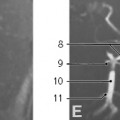
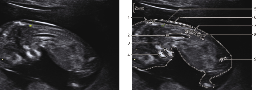
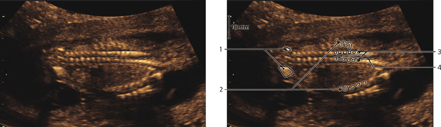
GA: 10w5d, CRL: 40 mm
- Amniotic cavity
- Leg
- Umbilical cord
- Placenta
- Maxilla
- Mandibula
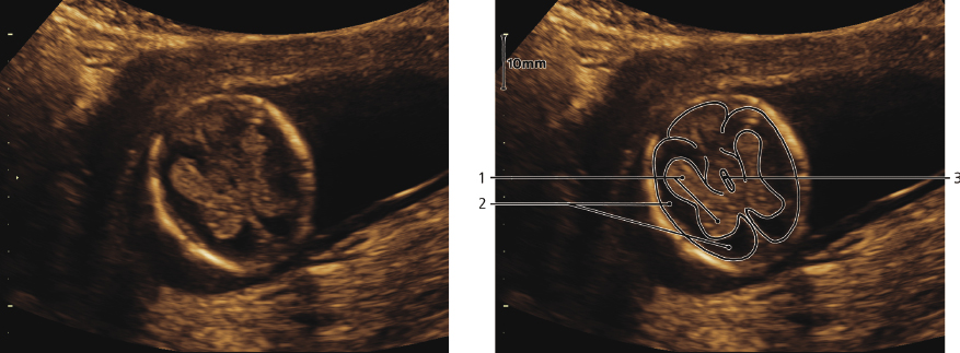
GA: 11w4d, head transverse
- Amniotic cavity
- Frontal bone
- Cerebral cortex
- Choroid plexus in lateral ventricle
- Temporal bone
- Parietal bone
- Occipital bone
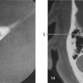
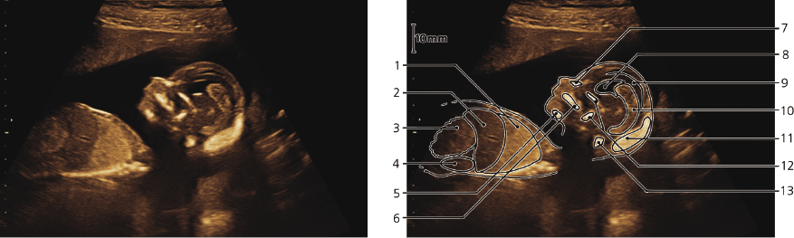
GA: 12w3d, neck, nuchal translucency, sagittal
- Occipital bone
- Aorta
- Mandibula
- Maxilla
- Nuchal translucency (“nuchal fold,” subcutaneous edema), NF 1.9 mm. (Normal for this GA is up to 2.4 mm)
- Reflection from skin
- Vertebral canal
- Vertebral bodies with ossification centers
- Ilium
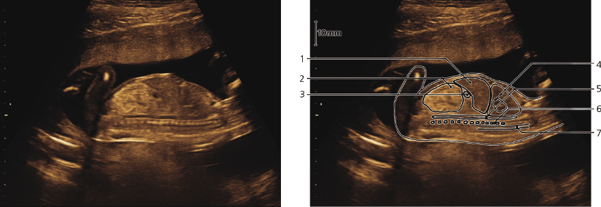
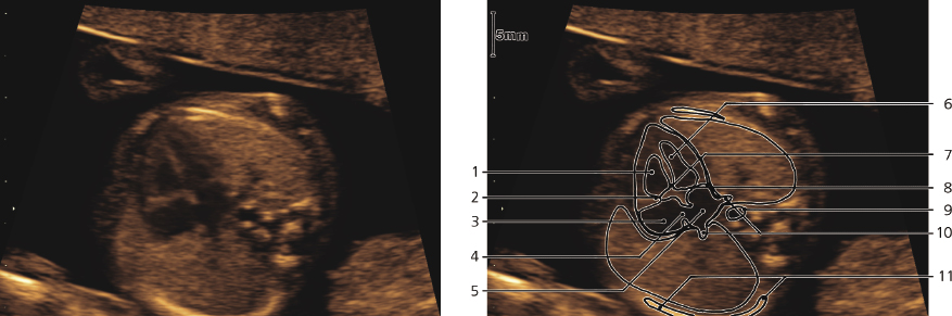
GA: 14w5d, head, sagittal
- Lung
- Liver
- Gut
- Kidney
- Mandibula
- Maxilla and palate
- Nasal bone
- Lateral ventricle
- Cerebral cortex
- Choroid plexus
- Occipital bone
- Sphenoid bone
- Atlas and axis
GA: 14w5d, brain, transverse
- Choroid plexus in lateral ventricle
- Cerebral cortex
- Third ventricle
GA: 14w6d, thorax, transverse
- Arm
- Heart
- Rib
- Ossification centers in vertebral arch
- Vertebral canal
- Ossification center in vertebral body
- Lungs
GA: 15w0d, spine, frontal
- Coxae
- Ribs
- Ossification centers in vertebral arch (thoracic)
- Vertebral canal
GA: 15w0d, spine, mid-sagittal
- Liver
- Gut
- Stomach
- Heart and lung
- Aorta
- Vertebral bodies
- Vertebral canal
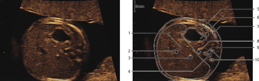
GA: 15w2d, four chamber view of heart
- Right ventricle
- Tricuspid valve
- Right atrium
- Oval foramen
- Left atrium
- Left ventricle
- Crux cordis
- Mitral valve
- Aorta
- Pulmonary veins
- Rib
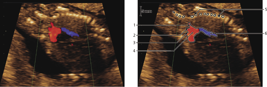
GA: 15w2d, aortic arch. Color-flow Doppler imaging
- Left subclavian artery
- Left common carotid artery
- Brachiocephalic trunk
- Aortic arch
- Vertebral column
- Descending aorta
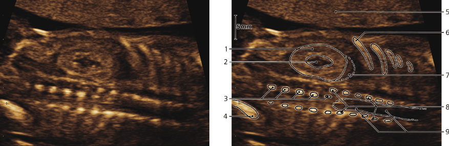
GA: 15w2d, upper abdomen, transverse
- Liver
- Umbilical vein
- Inferior caval vein
- Vertebral canal
- Spleen
- Stomach
- Rib
- Aorta
- Ossification center of vertebral body
- Ossification centers of vertebral arch
GA: 15w2d, spine, frontal. The shift between 3 and 8 is due to rotation
Only gold members can continue reading.
Log In or
Register to continue

Stay updated, free articles. Join our Telegram channel