However, matrix may range from purely lytic to purely sclerotic lesion
• Narrow zone of transition ± sclerotic margin
• Low to intermediate signal intensity on T1WI, heterogeneous on T2WI, STIR, variable enhancement
• Mild to marked increase in radionuclide uptake
Top Differential Diagnoses
• Aneurysmal bone cyst (ABC)
• Paget disease
• Osteoblastoma
• Osteosarcoma
• Tuberous sclerosis
Pathology
• Sporadic mutation in GNAS gene
Clinical Issues
• Growth disturbance, pathologic fracture
• Rarely undergoes sarcomatous transformation
Diagnostic Checklist
• Do not confuse with Paget disease on imaging
Paget disease thickens cortex and trabeculae
FD thins cortex and replaces trabeculae
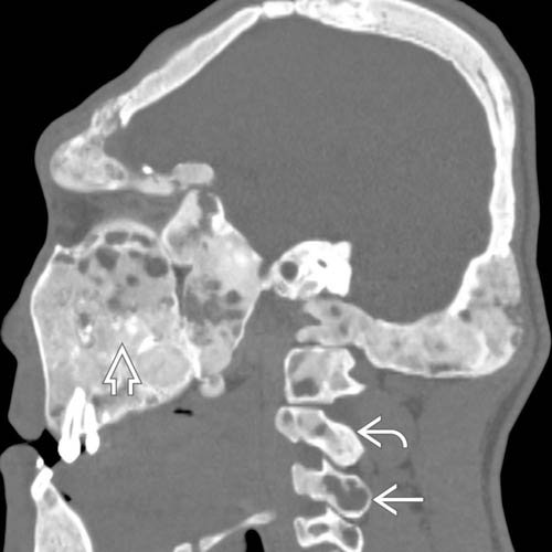
(Left) Sagittal bone CT shows severe polyostotic fibrous dysplasia (FD) involving skull, facial bones, and cervical spine. Some areas are “ground-glass” , others are purely lytic , and there are a few foci of calcified cartilage .
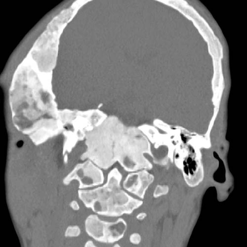
(Right) Coronal bone CT in the same patient shows loss of normal trabeculae in the majority of the included bones, replaced by fibrous dysplasia matrix. Thinning of tables of skull is a characteristic finding.
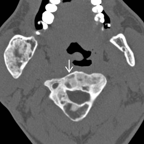
(Left) Axial bone CT shows variation in density in a single vertebra with nearly complete marrow replacement. This variability is common and should not raise suspicion for malignant degeneration.
(Right) Axial bone CT shows lytic FD in the posterior elements. The narrow zone of transition (sometimes sclerotic) helps to distinguish FD from more aggressive processes. Vertebral bodies tend to be less severely involved than the posterior elements.
TERMINOLOGY
Abbreviations
• Fibrous dysplasia (FD)
Definitions
• Monostotic: Single lesion
• Polyostotic: Multiple lesions, often associated with growth disturbances


 , others are purely lytic
  , and there are a few foci of calcified cartilage
  .
.
 in a single vertebra with nearly complete marrow replacement. This variability is common and should not raise suspicion for malignant degeneration.
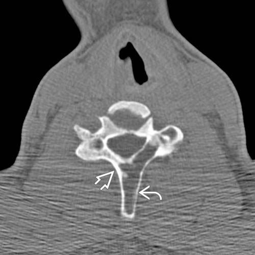
 in the posterior elements. The narrow zone of transition
  (sometimes sclerotic) helps to distinguish FD from more aggressive processes. Vertebral bodies tend to be less severely involved than the posterior elements.


