Presentation and Presenting Images
( ▶ Fig. 93.1, ▶ Fig. 93.2, ▶ Fig. 93.3, ▶ Fig. 93.4, ▶ Fig. 93.5, ▶ Fig. 93.6, ▶ Fig. 93.7)
A 51 year-old-female with a recent left breast biopsy at an outside institution consistent with ductal carcinoma in situ and invasive ductal carcinoma presents for diagnostic mammography and evaluation.
93.2 Key Images
( ▶ Fig. 93.8, ▶ Fig. 93.9, ▶ Fig. 93.10, ▶ Fig. 93.11, ▶ Fig. 93.12, ▶ Fig. 93.13, ▶ Fig. 93.14)
93.2.1 Breast Tissue Density
The breasts are heterogeneously dense, which may obscure small masses.
93.2.2 Imaging Findings
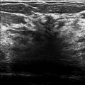
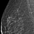
The imaging of the right breast is normal (not shown). The left breast demonstrates fine-linear branching calcifications, an associated tissue density, and a postbiopsy clip at the 3 o’clock location in the middle to posterior depth, 6 cm from the nipple. (circle) ( ▶ Fig. 93.1, ▶ Fig. 93.2, ▶ Fig. 93.3, ▶ Fig. 93.8, ▶ Fig. 93.9, ▶ Fig. 93.10). While tomosynthesis ( ▶ Fig. 93.4, ▶ Fig. 93.5, ▶ Fig. 93.11, ▶ Fig. 93.12) diminishes the effects of the tissue density which is consistent with a postbiopsy hematoma and allows the full extent of the calcifications to be seen, spot-magnification views ( ▶ Fig. 93.6, ▶ Fig. 93.7, ▶ Fig. 93.13, ▶ Fig. 93.14) are needed to evaluate the morphology of the calcifications.
93.3 BI-RADS Classification and Action
Category 5: Highly suggestive of malignancy (for new finding of fine-linear branching calcifications)
Category 6: Known biopsy-proven malignancy
93.4 Differential Diagnosis
Ductal carcinoma in situ: Fine-linear branching calcifications is a typical presentation of ductal carcinoma in situ. This morphology of calcifications warrants a BIRADS category 5.
Invasive ductal carcinoma: Typically invasive carcinomas do not present as calcifications.
Ductal carcinoma in situ and invasive ductal carcinoma: If the density was a mass lesion and not postbiopsy changes, this finding would be suspicious for ductal carcinoma in situ and invasive ductal carcinoma.
93.5 Essential Facts
The postbiopsy changes obscure findings on the conventional two-dimensional (2D) mammography. This masking is eliminated with DBT.
DBT allows the full extent of calcifications to be evaluated and measured, but it is unable to perform magnification views to characterize the calcifications. Characterization of the morphology of calcifications must be performed with 2D mammography.
Having an accurate size measurement helps to adequately stage the malignancy and to develop an appropriate treatment plan.
Although magnification views cannot be performed with DBT and calcifications may be on multiple slices, in this example, the calcifications were shown to be more extensive on tomosynthesis than seen on the conventional mammography.
93.6 Management and Digital Breast Tomosynthesis Principles
Tomosynthesis systems must support both conventional mammography and tomosynthesis because magnification views cannot be performed with tomosynthesis.
Spangler and colleagues (2011) found conventional mammography to be slightly more sensitive than DBT for the detection of calcifications, but, with improvements in processing algorithms and display, DBT could potentially improve.
Kopans and colleagues (2011) found that calcifications can be demonstrated with the same or better clarity on DBT as on conventional mammography, which allows the same and perhaps improved analysis of calcifications.
In this case DBT removed the postbiopsy changes that obscured the calcifications, allowing them to be visualized.
93.7 Further Reading
[1] Kopans D, Gavenonis S, Halpern E, Moore R. Calcifications in the breast and digital breast tomosynthesis. Breast J. 2011; 17(6): 638‐644 PubMed
[2] Spangler ML, Zuley ML, Sumkin JH, et al. Detection and classification of calcifications on digital breast tomosynthesis and 2D digital mammography: a comparison. AJR Am J Roentgenol. 2011; 196(2): 320‐324 PubMed

Fig. 93.1 Left cranciocaudal (LCC) mammogram.
Stay updated, free articles. Join our Telegram channel

Full access? Get Clinical Tree


