Presentation and Presenting Images
( ▶ Fig. 39.1, ▶ Fig. 39.2, ▶ Fig. 39.3, ▶ Fig. 39.4)
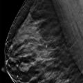
A 44-year-old female presents for routine screening mammography.
39.2 Key Images
39.2.1 Breast Tissue Density
The breasts are heterogeneously dense, which may obscure small masses.
39.2.2 Imaging Findings
There is a 1.2-cm focal asymmetry (circle in ▶ Fig. 39.5 and ▶ Fig. 39.6) in the upper outer quadrant of the left breast at the 2 o’clock location, 6 cm from the nipple. There are no suspicious findings on the right breast, and there are no prior mammograms of the left breast available for comparison.
39.3 BI-RADS Classification and Action
Category 0: Mammography: Incomplete. Need additional imaging evaluation and/or prior mammograms for comparison.
39.4 Diagnostic Images
39.4.1 Imaging Findings
At diagnostic imaging, only tomosynthesis was performed. The tomosynthesis movie demonstrated that the finding in the left breast is not a true lesion but is the result of overlapping breast parenchyma.
39.5 BI-RADS Classification and Action
Category 2: Benign
39.6 Differential Diagnosis
Summation artifact: This finding resolves on the diagnostic tomosynthesis movie, consistent with a pseudomass or summation artifact.
Asymmetry: This finding is seen on two views. An asymmetry is a one-view finding.
Developing asymmetry: There were no prior images for comparison so one cannot ascertain if this finding has increased in conspicuity or size over time.
39.7 Essential Facts
In this case the screening mammography was done without tomosynthesis. If a combination study with conventional mammography and tomosynthesis had been performed, this patient would not have been recalled for additional imaging because the finding would have resolved on tomosynthesis.
There are four types of asymmetries described in the ACR BI-RADS Atlas, 5th edition: asymmetry, focal asymmetry, developing asymmetry, and global asymmetry.
An asymmetry is a finding seen on only one mammographic view.
A focal asymmetry is a nonmass lesion visible on at least two mammographic views that occupies less than a quadrant.
A developing asymmetry is a focal asymmetry that is new, larger, or more conspicuous than noted previously. The risk of malignancy is 15% at screening mammography and 25% at diagnostic mammography, making this a suspicious finding.
A global asymmetry is a nonmass lesion visible on at least two mammographic views that occupies at least a quadrant.
39.8 Management and Digital Breast Tomosynthesis Principles
Tomosynthesis helps imagers to identify true mass lesions and to dismiss pseudomasses or summation artifacts.
The detection of lesions obscured by overlapping breast tissue is a limitation to conventional mammography.
Overlapping breast tissue can obscure cancers resulting in a missed cancer diagnosis. Also, overlapping breast tissue can create summation artifacts or pseudomasses resulting in a false-positive diagnosis and a biopsy of a benign finding.
Tomosynthesis reduces the effects of overlapping breast tissue, thus reducing callbacks and biopsies.
39.9 Further Reading
[1] Leung JWT, Sickles EA. Developing asymmetry identified on mammography: correlation with imaging outcome and pathologic findings. AJR Am J Roentgenol. 2007; 188(3): 667‐675 PubMed
[2] Sickles EA, D’Orsi CJ, Bassett LW, et al. ACR BI-RADS Mammography. In: ACR BI-RADS Atlas, 5th edition. Reston, VA: American College of Radiology; 2013

Fig. 39.1 Right craniocaudal (RCC) mammogram.
Related posts:


Stay updated, free articles. Join our Telegram channel

Full access? Get Clinical Tree


