and Zdeněk Fryšák1
(1)
Department of Internal Medicine III – Nephrology, Rheumatology and Endocrinology, Faculty of Medicine and Dentistry, Palacky University Olomouc and University Hospital Olomouc, Olomouc, Czech Republic
Keywords
Follicular thyroid carcinomaDistant metastasesPoorer survival rateDifferent ultrasound features14.1 Essential Facts
A follicular thyroid carcinoma (FTC) accounts for 10–32% of differentiated thyroid carcinomas (DTC); the rest is papillary thyroid carcinoma (PTC) [1].
In a large demographic study in the USA the second most common histologic type among all sex and racial/ethnic groups was FTC ranges 9–23%. Both PTC and FTC rates are consistently two to three times higher among females than males. Incidence rates tended to be higher among White than among Black subjects, and among White non-Hispanic subjects than among White Hispanic or Asian Pacific Islander subjects [2].
In iodine-deficient areas the relative rate of FTC tends to be even higher, up to 40% of the cases [3].
More recently, a decreased incidence of FTC has been reported. This decrease is probably due to more accurate histological diagnosis (exclusion of atypical follicular adenoma, identification of follicular variants of PTC) and also to iodine supplementation programs [3].
FTC has been classified as minimally invasive (MI-FTC), which has limited capsular and/or vascular invasion, and as widely invasive (WI-FTC), which has widespread infiltration of adjacent thyroid tissue and/or blood vessels. WI-FTC has larger primary tumors, and had a higher incidence of extrathyroidal extension, lymph node metastasis, and distant metastasis (50%) [1, 3].
LN involvement in FTC is uncommon, ranging from 0–10% [3].
DTC (PTC and FTC) have similarities in their clinically indolent behavior, management, and outcome. However, there are distinct differences between these two carcinomas: Patients with FTC tend to be older, present with more advanced disease, and have a poorer survival rate [1].
FTC has a higher risk of developing distant metastases (28.8%) and mortality (17.2%) compared with PTC (8.9% resp. 7.6%) [1].
The recurrences of PTC and FTC are most frequent at the extremes of age (<20 and >59 years), but their mortality rates successively increased in patients aged >40 years [4].
The overall 5-year relative survival rate for PTC and FTC is greater than 90%; FTC, therefore, has a poorer prognosis compared to PTC. The mortality rate ranges from 5–15% even if the disease is confined to the thyroid at the time of diagnosis [5].
For a more detailed comparison of both cancers, see Sect. 15.2 of Chap. 15.
Hürthle cell carcinoma (HCC) has generally been considered to be a variant of FTC. HCC occurs in an older age group, is more aggressive with distant metastasis, and has a poorer prognosis than non-HCC [3]. HCC accounts for 3–5% of all thyroid cancers [7, 8]. In a study by Kushchayeva et al. [9] the 10-year disease-free interval was 75% for FTC and 40.5% for HCC and the 10-year cause-specific mortality was 20% for FTC and 51% for HCC patients [9]. Increasing age, male sex, and increasing tumor size substantially decrease survival of patients with HCC [7].
14.2 US Features of FTC According to the 2015 ATA Guidelines [6]
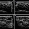
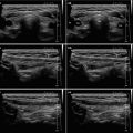
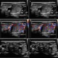
For FTC, apply the same 2015 ATA criteria as for others suspected nodes (Fig. 14.1aa).

Fig. 14.1


(aa) A 76-year-old woman with two follicular thyroid carcinomas (FTCs) in the LL—micro FTC, size 9 × 9 × 8 mm, volume 0.5 mL and small FTC, size 19 × 15 × 12 mm, volume 2 mL: micro FTC (blank arrowheads)—“taller-than-wide” shape; small FTC (arrowheads)—round shape; both with the same US appearance—solid; inhomogeneous structure; mostly hypoechoic; smooth margin; LL 8 mL; transverse. (bb) Detail of two FTCs, CFDS: micro FTC (blank arrowheads)—minimal vascularity at periphery, pattern 0; small FTC (arrowheads)—increased central and peripheral vascularity, pattern II; transverse. (cc) Detail of small FTC (arrowheads): ovoid shape; longitudinal. (dd) Detail of small FTC (arrowheads), CFDS: increased central and peripheral vascularity, pattern II; longitudinal. (ee) Detail of micro FTC (blank arrowheads): “taller-than-wide” shape; longitudinal. (ff) Detail of micro FTC (blank arrowheads), CFDS: minimal peripheral vascularity, pattern 0; longitudinal
Related posts:
Stay updated, free articles. Join our Telegram channel
Full access? Get Clinical Tree
