Chapter 6 Forearm, elbow and humerus
Forearm (radius and ulna)
This region of the upper limb most usually presents for imaging as a result of trauma. The Colles’ fracture is the most usual finding after trauma to radius and ulna; this is outlined in Chapter 5 (section on the wrist). Other fractures of these bones are much rarer. The Galleazzi fracture is more serious than the Colles’, being a fracture of the distal portion of the radius accompanied by subluxation or dislocation of the distal radioulnar joint. The Monteggia fracture, conversely, is a fracture of the ulna accompanied by dislocation of the radius proximally.1
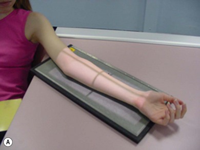
Anteroposterior (AP) forearm (Fig. 6.1A,B)
Positioning
• The patient is seated with the affected side next to the table; lead rubber is applied to the waist
• The arm is extended at the elbow, abducted away from the trunk and externally rotated until the hand lies in supination
• The posterior aspect of the forearm is placed in contact with the IR, to include elbow and wrist joints
• The joints must lie in the same plane
• The humeral epicondyles and radial and ulnar styloid processes are equidistant from the IR
• The head is turned away from the shoulder of the side under examination, aiming to reduce scattered radiation to the lenses of the eyes and thyroid
Criteria for assessing image quality
• Wrist and elbow joints, radius, ulna and soft tissue outline of the forearm are demonstrated
• Partial superimposition of the radius and ulna at proximal and distal ends, with separation of the shafts. Radial tubercle should overlap the cortex of the ulnar shaft, but no further
• Humeral epicondyles equidistant from coronoid and olecranon fossae
• Radial styloid process seen on the lateral aspects of this bone
• Ulnar styloid process is shown in profile distally in the middle of the head of ulna
• Sharp image demonstrating soft tissue margins of the forearm, bony cortex and trabeculae. Adequate penetration to demonstrate overlap of olecranon over distal humerus while showing trabecular detail over shafts of radius and ulna
| Common errors | Possible reasons |
|---|---|
| Radius cleared from ulna at the proximal end; radial head also shown clear | Externally rotated arm |
| Radial tubercle superimposed over shaft of ulna | Internally rotated arm |
| Shafts of radius and ulna show adequate contrast and density but elbow is ‘thin’, underpenetrated and shows poor contrast or bony detail | Inadequate kVp selected |
| Elbow joint shows adequate contrast and density but shafts of radius and ulna are dark, showing poor contrast and bony detail | Selected kVp too high |
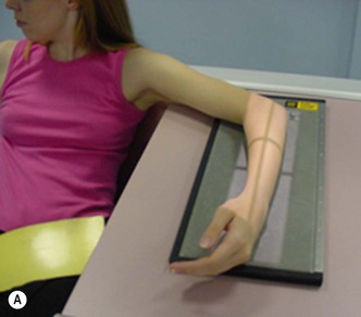
Lateral forearm (Fig. 6.2A,B)
Positioning
• The patient is seated with the affected side next to the table; lead rubber is applied to the waist
• The arm is flexed at the elbow, abducted away from the trunk and internally rotated at the wrist
• The medial aspect of the forearm is placed in contact with the IR, to include elbow and wrist joints
• The shoulder, elbow and wrist joints must lie in the same plane
• The humeral epicondyles are superimposed, as are the radial and ulnar styloid processes. Ensuring the shoulder lies in the same plane as the wrist and elbow will help facilitate this
• The head is turned away from the shoulder of the side under examination, aiming to reduce scattered radiation to the lenses of the eyes and thyroid
Criteria for assessing image quality
• The wrist and elbow joints, radius, ulna and soft tissue outline of the forearm are demonstrated
• Superimposition of posterior portion of radial head over coronoid process of ulna; superimposition of distal radius and ulna
• Shaft of the radius is seen anterior to that of the ulna
• There will be some superimposition of trochlea and capitulum of humerus. However, it may be unrealistic to expect to see full superimposition of these structures as the obliquity of the beam at its periphery is likely to pass through the elbow at around 3–4°
• Sharp image demonstrating soft tissue margins of the forearm, bony cortex and trabeculae. Adequate penetration to demonstrate overlap of radial head over the olecranon and distal radius over ulna, while showing trabecular detail over the shafts of the radius and ulna
| Common errors | Possible reasons |
|---|---|
| Distal radius seen anteriorly in relationship to ulna | Wrist is medially rotated |
| Distal radius seen posteriorly in relationship to ulna; shafts of radius and ulna superimposed along most of their length | Wrist and elbow are externally rotated; this usually only occurs when the humerus does not lie in the same plane as the forearm and the shoulder lies above the table-top |
Elbow
The supracondylar fracture of the humerus has many implications for the future of the patient’s arm. The vasculature of the arm can be damaged, or existing damage can be exacerbated, by forced extension of the elbow joint; this can cause an ischaemic state in the lower arm resulting in paralysis of the hand and forearm and, long term, in what is known as a Volkmann’s ischaemic contracture. It is therefore essential that the radiographer undertakes modified projections of the elbow which cannot be extended; these are outlined in Chapter 25 on accident and emergency (A&E) radiography.
For all projections of the elbow the IR is placed horizontal unless otherwise specified.
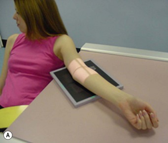
AP elbow (Fig. 6.3A,B)
Positioning
• The patient is seated with the affected side next to the table; lead rubber is applied to the waist
• The arm is extended at the elbow, abducted away from the trunk and externally rotated until the hand lies in supination
• The posterior aspect of the elbow is placed in contact with the IR
• The wrist, elbow and shoulder joints must lie in the same plane
• The humeral epicondyles are equidistant from the IR
• The head is turned away from the shoulder of the side under examination, aiming to reduce scattered radiation to the lenses of the eyes and thyroid
Criteria for assessing image quality
• The proximal radius and ulna, elbow joint, distal shaft of the humerus and soft tissue outlines surrounding the elbow joint demonstrated
• Partial superimposition of the radius and ulna at the proximal end (0.6 cm of radial head superimposed over ulna).2 Radial tuberosity should overlap the cortex of the ulnar shaft, but no further
• Humeral epicondyles equidistant from the coronoid and olecranon fossae
• Sharp image demonstrating soft tissue margins around the elbow, bony cortex and trabeculae. Adequate penetration to demonstrate overlap of olecranon over distal humerus
| Common errors | Possible reasons |
|---|---|
| Radius cleared from ulna; radial head also shown clear | Elbow is externally rotated |
| Radial head superimposed more than 0.6 cm over shaft of ulna | Internally rotated elbow |
| Radial head fully superimposed over ulna; distance between humeral epicondyles seems narrow | Hand may be in pronation rather than supination |
| Joint space between capitulum and radial head is closed; long axes of radius and ulna travel obliquely towards the lateral aspect of the arm away from the joint | Arm not fully extended at the elbow |
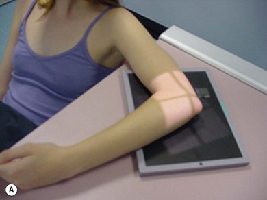
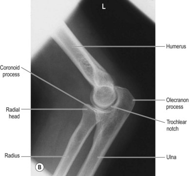
Lateral elbow (Fig. 6.4A,B)
Positioning
• The patient is seated with the affected side next to the table; lead rubber is applied to the waist
• The arm is abducted from the trunk, internally rotated and flexed 90° at the elbow
• The wrist is externally rotated until the radial and ulnar styloid processes are superimposed
• The medial aspect of the elbow is placed in contact with the IR
• The shoulder, elbow and wrist joints must lie in the same plane
• The humeral epicondyles are superimposed. Ensuring the shoulder lies in the same plane as the wrist and elbow will help facilitate this more easily
• The head is turned away from the shoulder of the side under examination, aiming to reduce scattered radiation to the lenses of the eyes and thyroid
Criteria for assessing image quality
• The proximal radius and ulna, elbow joint, distal shaft of the humerus and soft tissue outlines surrounding the elbow joint are demonstrated
• Superimposition of surfaces of trochlea and capitulum, with the posterior portion of the radial head shown over the coronoid process of ulna. Evidence of joint space of the elbow seen
• Shaft of the radius is seen anterior to that of the ulna
• Sharp image demonstrating soft tissue margins around the elbow, bony cortex and trabeculae. Adequate penetration to demonstrate overlap of radial head over the olecranon and superimposed epicondyles. Exposure factors must ensure that the anterior and supinator fat pads are shown in contrast with the surrounding soft tissue (the posterior fat pad will only be demonstrated if there is bony injury)
The importance of optimum exposure factor selection cannot be emphasised enough, especially in the case of the elbow radiograph requested after trauma. Information on both bone and soft tissue becomes even more vital in trauma cases. This is because personnel assessing and/or reporting on the radiograph need to inspect the image for evidence of the ‘fat pad sign’, an indication of presence of abnormal fluid (usually blood) outside the elbow’s joint capsule. This sign suggests bony damage, often supracondylar or radial head fractures, which may or may not be evident on the radiograph. When significant trauma causes displacement of the pads there will be an appearance similar to a downturned rose thorn (seen as darker than the surrounding soft tissue) anterior and/or posterior to the distal humerus, just above the epicondyles. The normal positions of the fat pads are: supinator fat pad seen along the anterior aspect of the humerus; anterior fat pad seen anterior to the distal portion of humerus just above the coronoid fossa; the posterior fat pad is positioned within the olecranon fossa posteriorly.1
Flexion of the joint also affects fat pad appearance in the lateral elbow projection. Flexion <90° causes the olecranon to move towards the olecranon fossa, thereby displacing the posterior pad superiorly to a position which may be visible on the lateral radiograph. This may potentially mimic appearances suggestive of trauma and thus affect radiological comment.2
Stay updated, free articles. Join our Telegram channel

Full access? Get Clinical Tree


