99mTc-BRIDA (mins)
Hepatic Tmax
12
Hepatic T ½
15–20
Principal bile duct visualization
5–20
Gallbladder visualization
10–40
Tracer appearing in the bowel
15–45
19.2 Teaching Cases
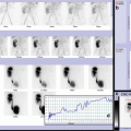
19.2.1 Case 19.1 Hepatobiliary Scintigraphy in Diagnostic Workup of Recurrent Pancreatitis
A 9-year-old girl with Crohn’s disease diagnosed at the age of 5 years. Subsequently, she had recurrent episodes of acute pancreatitis secondary to papillary stenosis and underwent surgical treatment. Three years after treatment, she had relapsed episodes of pancreatitis. Ultrasonography and magnetic resonance imaging showed severe chronic pancreatitis with pancreatic ducts dilatation, splenomegaly, and increased gallbladder wall thickening without biliary ducts dilatation. A hepatobiliary scintigraphy was performed to evaluate liver function and biliary system excretion before further surgical treatment. A normal hepatic function was detected with regular parenchymal transit and transient tracer stasis in the gallbladder (as for gallbladder atonia).
Radiological imaging plays a major role in the diagnostic workup of children with hepatobiliary or pancreatic diseases, while hepatobiliary scintigraphy is performed in selected cases to achieve functional and biliary drainage data (Fig. 19.1).
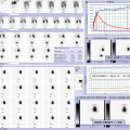
19.2.2 Case 19.2 Biliary Atresia in a Newborn with Prolonged Neonatal Cholestasis
A 4-week-old boy with tetralogy of Fallot, cholestatic jaundice, and pale stool. Laboratory examination showed elevated total bilirubin with an increased conjugated portion, slightly elevated gGT. Hepatobiliary scintigraphy reported, “no drainage” of the radiolabeled tracer in the small bowel within 24 h as in extrahepatic biliary atresia (BA) patients. A percutaneous liver biopsy showed obstructive cholestasis. Diagnosis of BA was confirmed at 2 months of age by intraoperative cholangiography during Kasai portoenterostomy. Diagnosis of BA and performance of a hepatoportoenterostomy (Kasai Procedure) by 8–10 weeks of age are optimal for transplant-free survival beyond early childhood (Fig. 19.2).
19.2.3 Case 19.3 Late Diagnosis of Biliary Atresia with Secondary Liver Impairment
A 6-month-old boy referred to our hospital presenting with chronic cholestatic jaundice, high bilirubin level, splenomegaly, and acholic stool. Abdominal ultrasound and hepatobiliary scintigraphy were promptly performed, establishing the diagnosis of biliary atresia. Biliary atresia (BA) is universally fatal if untreated and is the single most common cause of liver disease leading to liver transplantation in children. In BA, bile flow from the liver to the gallbladder is blocked, and bile trapped inside the liver progressively causes damage and scarring of the liver cells (cirrhosis) and liver failure. Hepatobiliary scintigraphy revealed liver impairment in this patient due to late diagnosis of BA, and liver transplantation was necessary after few months (Fig. 19.3).
19.2.4 Case 19.4 Progressive Liver Failure After Performance of Hepatoportoenterostomy (Kasai Procedure) with Scintigraphic Finding of Bile Ascites
A 5-month-old boy diagnosed with biliary atresia at 4 weeks of age. At 2 months of age, a Kasai portoenterostomy was performed. He did not afford the restoration of the bile flow after the procedure and developed cirrhosis and ascites. In addition to radiological imaging, a hepatobiliary scintigraphy was performed showing liver disease progression with low hepatic function and biliary ascites (confirmed by ascitic fluid analysis). He underwent a liver transplant after 4 months.
Infants with biliary atresia who undergo a successful Kasai procedure regain good health and no longer have jaundice or major liver problems. When Kasai procedure is not successful, infants usually need a liver transplant within 1–2 years. Even after a successful surgery, most infants with biliary atresia could slowly develop cirrhosis over the years requiring a liver transplant by adulthood (Fig. 19.4).
19.2.5 Case 19.5 Diagnostic Imaging for Differential Diagnosis Between Cystic Biliary Atresia and a Congenital Choledochal Malformation
A 5-week-old female presented with jaundice and acholic stools to our center. She underwent a complete hepatology assessment that ruled out metabolic or infectious causes of her cholestasis. Ultrasound scan of the abdomen showed evidence of a choledochal cyst and sonographic signs suspicious of biliary atresia. Therefore, hepatobiliary scintigraphy was performed, and “no draining” of the radiolabed tracer in the small bowel within 24 h was seen, as in keeping with extrahepatic biliary atresia. Soon after the nuclear imaging, an intraoperative cholangiography confirmed the diagnosis of biliary atresia, and a portoenterostomy was done. In a small number of cases, biliary atresia may be associated with choledochal cyst, and preoperative diagnostic workup is crucial to select the right surgical approach (Fig. 19.5).
19.2.6 Case 19.6 Diagnostic Workup in a Case of a Choledochal Cyst
A 1-month-old boy, with no picture of cholestasis, underwent an abdominal ultrasound scan during the workup for intestinal motility disorders. Ultrasound abdominal scan revealed the presence of biliary dilatation, and hepatobiliary scintigraphy was performed to rule out the less likelihood hypothesis of a biliary atresia associated with choledochal cyst. Nuclear medicine imaging revealed a normal liver function and further clarified the biliary excretion, showing an early photon-deficient area in the porta hepatis with biliary tracer filling on late images (as for choledochal cysts); physiological gut activity was detected excluding biliary atresia association. A diagnosis of choledochal cyst was made and surgery planned (Fig. 19.6).
19.2.7 Case 19.7 Hepatobiliary Scintigraphy in Diagnostic Workup of Choledochal Cyst Complicated by Perforation of Bile Duct
A 2-month-old boy came to Emergency with clinical presentation of ascites and acholic stools. Previous history was uneventful with physiological neonatal jaundice and a prenatal ultrasonography referred as normal. Radiological imaging showed evidence of a large choledochal cyst, and a drainage tube was placed to evacuate and characterize the nature of abdominal fluid. By macroscopic analysis, the abdominal fluid had biliary color; chemical analysis confirmed high bilirubin level (12.9 mg/dL), whereas blood bilirubin level was quite normal (1.4 mg/dL), and all cultures performed were negative. These findings were indicative of perforation of bile duct that is an uncommon but possible event, consequent to different causes (trauma, ischemia, necrotizing enterocolitis, congenital malformation of bile ducts, or spontaneous breakage). Hepatobiliary scintigraphy was performed to achieve a complete evaluation of biliary drainage in relation to malformation of biliary tree complicated by abdominal leak (Fig. 19.7).
19.2.8 Case 19.8 Hepatobiliary Scintigraphy for Differential Diagnosis Between Hepatic Cyst and Biliary Origin Lesion
A 3-month-old boy with prenatal diagnosis of hepatic cyst (maximum axial diameter of 13 mm) in the VIII segment. Ultrasound postnatal abdominal scan confirmed the presence of hepatic lesion with significant increased dimension (maximum axial diameter of 2.8 cm). The newborn presented good clinical condition even if jaundice with high indirect bilirubin component was present (6.7 vs. 0.28 mg/dL total vs direct respectively). Two further abdominal ultrasounds were performed showing a dimensional increase of hepatic lesion (up to 4.3 × 3 × 3 cm) corresponding to the hepatic segments VIII, V, and IV. No evidence of pale stools was reported. Alpha-1 fetoprotein level was normal for age. Hepatobiliary scintigraphy was performed to rule out the biliary origin of the lesion by biliary drainage evaluation, but no evidence of biliary tracer uptake was observed corresponding to the hepatic lesion detected by ultrasound scan.
In the following years, the child remained asymptomatic, and a strict sonographic follow-up revealed stable morphological features of the lesion, excluding (up to the last clinical control) surgical treatment indications (Fig. 19.8).
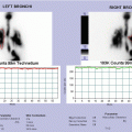
19.2.9 Case 19.9 Hepatobiliary Scintigraphy in Liver Disease Assessment of Children with Cystic Fibrosis and Signs of Cirrhotic Evolution
A 9-year-old boy with cystic fibrosis had a full hepatological review for assessment of his liver disease. The child was in poor clinical conditions presenting severe cirrhosis associated with chronic liver failure with signs of portal hypertension, protein synthesis defects, and mixed hyperbilirubinemia. Hepatobiliary scintigraphy was part of the assessment, and the result was in keeping with the previous laboratory, clinical, and radiological evaluations of severe chronic liver disease with signs of cirrhotic evolution. The patient was scheduled for liver transplantation (Fig. 19.9).
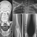
19.2.10 Case 19.10 Hepatobiliary Scintigraphy in Biliary Drainage Assessment of Children with Hepatic Angiomatosis and Intractable Pruritus
A 13-year-old girl with cutaneous and hepatic angiomatosis at birth. By interferon treatment, she had regression of dermal component but persistence of hepatic impairment. The child was clinically followed up with periodical blood analysis and ultrasonography scans until the age of 11 when itching appeared without any response to medical treatment. Hepatobiliary scintigraphy was performed showing moderately reduced liver function and biliary stasis secondary to intrahepatic bile ducts compression and distortion by vascular malformation. The girl underwent liver transplantation with an uneventful follow-up. Biopsy performed at 5 years after transplantation did not show any significant abnormalities Fig. 19.10.
19.2.11 Case 19.11 Hepatobiliary Scintigraphy in Biliary Drainage Assessment of Children with Congenital Bile Duct Paucity and Relapse of Intractable Pruritus After Internal Biliary Diversion Treatment
A 12-year-old girl with progressive intrahepatic cholestasis type II (PFIC II, secondary to congenital bile duct paucity detected by biopsy) underwent internal biliary diversion (direct biliary–enteric bypass) because of intractable pruritus after all medications failed to control the symptom. Soon after surgery, she clinically improved; however, after 12 months, pruritus was intense as much as before. A hepatobiliary scintigraphy was performed to investigate biliary drainage and in particular gallbladder biliary excretion into the diversion (bowel loop) to rule out obstruction as surgical complications (Fig. 19.11).
19.2.12 Case 19.12 Hepatobiliary Scintigraphy in Biliary Drainage Assessment of Children Underwent Liver Transplantation with Suspect of Biliary Stasis
A 2-year-old boy with liver transplantation for cryptogenic cirrhosis. After liver transplantation, he had constant increase of cholestasis indices with suspect of biliary stasis. Hepatobiliary scintigraphy was performed to rule out obstruction as surgical complications by biliary drainage assessment (Fig. 19.12).
Stay updated, free articles. Join our Telegram channel

Full access? Get Clinical Tree


