Fig. 17.1
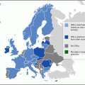
(a, b) By qualitative imaging and time/activity curves evaluation, a normal swallowing is observed with a regular passage of the radioactive bolus through the esophagus. No evidence of pulmonary aspiration episodes is detected during the registration. Some episodes of gastroesophageal reflux are evident, also with a small amount of labeled tracer

Fig. 17.2
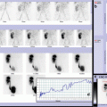
(a, b) Radionuclide salivagram shows massive and bilateral salivary aspiration in tracheal cannula and both lungs. A large amount of administered labeled bolus is visualized in the respiratory airways (proximal and distal), with evidence of mild tracer activity in the lower tract of esophagus and in the stomach. Partial clearance of aspirated tracer is evident at 4th and 23rd minute of registration in relation to tracheobronchial aspiration (necessary for breathing difficulties associated with desaturation). Eventually caught episodes or tracheobronchial aspiration procedure must be annotated and considered for a correct evaluation of aspirated clearance mechanism. This finding is considered as massive pulmonary aspiration

Fig. 17.3
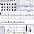
(a, b) After swallowing, fast progression of salivary secretion is evident in upper esophageal tract, and most of the salivary secretion passes directly into the trachea and in the right bronchus. Tracer penetration is subsequently evident into the left bronchus and in the distal airways of both lungs. Qualitative and T/A curves analyses show a progressive increased concentration of aspirated tracer (especially on the right). Low amount of labeled saliva is evident in the lower esophagus and in the stomach

Fig. 17.4


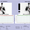
(a, b) After swallowing, progression of salivary secretion is evident in the esophagus showing a significant deviation of its course (secondary to a severe scoliosis) and a persistent tracer stasis in the distal tract due to a tight gastric fundoplication. A weak signal activity is documented in the right lung field, as for micro-aspiration scintigraphic sign
Stay updated, free articles. Join our Telegram channel

Full access? Get Clinical Tree


