Fig. 16.1
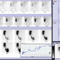
Scintigraphy is performed using seriated images (at 5′, 15′, 30′, 45′, and 60′ after the intravenous administration of Tc99m pertechnetate) in anterior view, with the child in supine position. In case of uncooperative children, it is also helpful turning the child in prone position, setting the head of gamma camera rotated below the patient. In the first image is evident a physiological tracer distribution in the stomach, cardiac blood pool, liver, spleen, and bladder; urinary tracer excretion could also be seen in the renal pelvis and ureters. There is no ectopic site of focal tracer accumulation suspicious for Meckel’s diverticulum. A normal rapid gastric uptake followed by a progressive increased and a physiological intestinal tracer progression appreciable during images acquisition is evident. In early phase, a slight and diffuse tracer uptake is detectable in lower abdomen, as for nonspecific intestinal inflammation
16.2.2 Case 16.2 A Scintigraphic Pattern Indicative of Meckel’s Diverticulum in Child with Rectal Bleeding
A 15-month-old male child was admitted to our emergency department after several episodes of rectal bleeding and a moderate anemia. An abdominal scintigraphy with 99m Tc-pertechnetate was performed showing an oval area of pathological pooling of the marker at the level of the middle-right abdomen, and a Meckel’s diverticulum was suspected. The patient underwent a laparoscopic-assisted resection of Meckel’s diverticulum, and the histology confirmed the presence of ectopic gastric mucosa at the level of the bowel resection. The 99mTc pertechnetate abdominal scintigraphy confirmed also in this case its role in the diagnosis of Meckel’s diverticulum (Fig.16.2).


Fig. 16.2
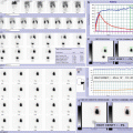
(a) Scintigraphy is performed using seriated images (at 5′, 15′, 30′, 45′, and 60′ after the intravenous administration of Tc99m-pertechnetate). In the first image, an abnormal area of focal uptake can be seen in the middle-right abdomen that fades at 15 min but reveals a following tracer increasing concentration up to the last image. An early and transient uptake in right abdomen could be a false-positive result caused by urinary retention activity in the right kidney (sometimes in lower position than usual), but following increasing concentration and anterior localization in lateral view are indicative of scintigraphic pattern of Meckel’s diverticulum. Lateral image should be performed as soon as a suspicious focal uptake is detected to avoid subsequent overlap with physiological tracer progression. (b) Intraoperative image of Meckel’s diverticulum during laparoscopic-assisted resection. Histology confirmed the presence of ectopic gastric mucosa at the level of the surgical specimen
16.2.3 Case 16.3 A Scintigraphic Pattern Indicative of Meckel’s Diverticulum After Intravenous Ranitidine Administration
An 8-year-old male child was referred to our unit after three episodes of melena. The esophagogastroduodenoscopy was negative, and the colonoscopy demonstrated a slight, nonspecific inflammation of the right colon. A previous standard 99m Tc abdominal scintigraphy performed in another institution was negative for radiomarker pooling. Due to the persistence of episodes of melena, despite the administration of mesalamine, we decided to repeat the Tc-99m abdominal scintigraphy. To improve the sensitivity, intravenous ranitidine was previously administrated, and an abnormal oval pooling of the radiotracer in the right abdomen was demonstrated suspicious for Meckel’s diverticulum. The patient underwent a minimally invasive laparotomic resection of the diverticulum, confirmed by the histopathology of the specimen. In this case, the previous administration of H2 inhibitors improved the sensitivity of the Tc99m scintigraphy and helped to confirm the clinical suspect of a Meckel’s diverticulum (Fig.16.3).


Fig. 16.3
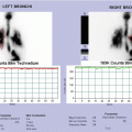
Tc99m pertechnetate abdominal scintigraphy is performed after previous intravenous ranitidine administration. Ranitidine premedication allows to inhibit acid secretion by parietal cells, thus limiting release of Tc99m pertechnetate by the mucosal cells and improving the sensitivity of the Meckel scan. The study shows an intense focus of increased tracer uptake in the right upper abdominal quadrant, seen simultaneously to stomach visualization and persisting up to the last static acquisition. In the first image, physiological tracer uptake in the right renal pelvis is evident in the region above focal area of intense pertechnetate concentration. A lateral image confirms anterior localization of focal abnormal uptake within intestine. This finding is strongly suspicious for Meckel’s diverticulum even if nonspecific inflammation of the right colon (especially when ulcerations are evident as detected by colonoscopy) could be the cause of false positive
16.2.4 Case 16.4 Characteristic Scintigraphic Pattern of Meckel’s Diverticulum with Progressive Increasing Focal Uptake
A 14-year-old boy was admitted to our emergency department for syncopal episode and 2 days long history of abdominal pain and black stool, suspected for melena. The patient was sick and pale; no history of prematurity and previous NSAIDs consumption. Blood tests showed worsening anemia that was treated with blood transfusion; abdominal ultrasound was normal. An upper gastrointestinal endoscopy excluded esophagitis, gastritis and duodenitis, and active bleeding. A 99mTc-pertechnetate scintigraphy detected a focal accumulation of radionuclide near the iliac bifurcation. In order to confirm the scintigraphic suspect of Meckel’s diverticulum, a minimally invasive laparotomic approach was performed with resection of the diverticulum. The postoperative period and follow-up were without complications (Fig.16.4).


Fig. 16.4
99m Tc-pertechnetate abdominal scintigraphy reveals a minimal focus of mild abnormal tracer uptake corresponding to inferior lower quadrants (at 5 min) with progressive tracer increasing concentration, up to the last image. This scintigraphic pattern is characteristic of Meckel’s diverticulum, confirmed by surgical and histological findings
16.2.5 Case 16.5 Scintigraphic Findings of Meckel’s Diverticulum with Variable Positions of Focal Tracer Uptake
A 6-year-old male child was admitted in our hospital for a Kawasaki syndrome, without cardiac complications. During hospitalization, the patient presented blood and mucus into the stool, progressive anemia, absence of abdominal pain. The routine laboratory tests and the levels of fecal calprotectin and anti-Saccharomyces cerevisiae antibody (ASCA) were normal. A colonoscopy excluded active bleedings. A 99mTc pertechnetate scintigraphy documented a focal accumulation of the radionuclide near the terminal ileum. On the basis of the scintigraphic findings, a surgical exploration was performed showing a Meckel’s diverticulum 30 cm above the ileocecal valve. Presence of ectopic gastric mucosa into the specimen was histologically confirmed. No complications occurred in the postoperative period and during the follow-up (Fig.16.5).


Fig. 16.5
99mTc-pertechnetate abdominal scintigraphy reveals an abnormal focus of tracer uptake corresponding to lower quadrants. This finding changes position in relation to bowel loop movements showing partial wash out at 30 and 70 min, and increasing concentration on the following images. A prolonged imaging (delayed image at 3 h) with empty bladder is helpful to confirm focal abdominal uptake strongly suggestive of Meckel’s diverticulum. Surgical resection of Meckel’s diverticulum is performed confirming the presence of ectopic gastric mucosa in the resected intestinal segment. However, a similar scintigraphic pattern could be due to gastrointestinal bleeding nonrelated to Meckel’s diverticulum or presence of severe bowel inflammation
16.2.6 Case 16.6 Diagnostic Imaging of Meckel’s Diverticulum: Scintigraphic and Radiological Findings
An 11-year-old boy admitted in our pediatric emergency department after an onset of abdominal pain associated to diarrhea with blood and mucus into the stool. His past clinical history was characterized by recurrent abdominal pain and one episode of subacute proximal small bowel obstruction at the age of 5. During hospitalization, laboratory tests were normal, but the level of fecal calprotectin was increased. An upper gastrointestinal series showed a suspect of Meckel’s diverticulum. To confirm the diagnosis and identify ectopic gastric mucosa, a 99mTc pertechnetate scintigraphy was performed. The scintigraphic pattern was characterized by a round focal accumulation of pertechnetate below iliac bifurcation. During a laparoscopic-assisted exploration, a Meckel’s diverticulum with a thick inflammatory band linked to the appendix was found. Resection of the diverticulum and appendectomy were performed without complications (Fig.16.6).


Fig. 16.6
(a) Upper GI series documented an image at the level of distal ileum that strongly suggested a Meckel’s diverticulum. (b) Tc99mpertechnetate abdominal scintigraphy is performed few days after radiological examination to ensure the absence of barium in the abdomen. Residual contrast in bowel from previous barium studies may hinder scintigraphic detection of ectopic gastric mucosa with false-negative results. In early phase, a slight and diffuse tracer uptake is evident in the abdomen corresponding to the lower side, as for nonspecific intestinal inflammation. Since image performed at 15 min, a focal area of tracer uptake is evident corresponding to lower quadrants, in median side, with following increasing concentration up to the last image. Approximately, 57 % of Meckel’s diverticula contain ectopic gastric mucosa which actively secretes hydrochloric acid responsible for mucosal ulcerations within the diverticulum and unprotected wall of adjacent ileum (causing gastrointestinal bleeding). This scintigraphic result reveals uptake of radiopertechnetate by functioning ectopic gastric mucosa in the diverticulum, detected by radiological imaging; for such reason, it is important to perform scintigraphic evaluation in acute or subacute bleeding phase
16.2.7 Case 16.7 False-Positive Scintigraphic Findings due to Severe Ulcerative Bowel Inflammation
A 2-year-old male patient was admitted in our hospital after an episode of intestinal bleeding. Laboratory tests documented: Hb 13.2 g/dL, WBC 11330/mmc; negative stool cultures. An abdominal ultrasonography showed bowel distension with liquid into the intestinal loops; no lesions in liver, pancreas, and spleen. Due to persistence of hematochezia, a 99mTc pertechnetate scintigraphy was performed with a suspect of ectopic gastric mucosa. Whereupon, a surgical exploration excluded a Meckel’s diverticulum. In order to complete the intestinal assessment, a colonoscopy showing severe ulcerative pancolitis was performed. At first, the child was treated with steroids and immunosuppressant drugs, but then a total colectomy was necessary because of the aggressive course of the disease (Fig.16.7).

