Gastrointestinal Malignancies


Gastrointestinal Malignancies


Koichi Tokuuye

Hirotoshi Kato

Shigeru Yamada

Tadashi Kamada

Yasuyuki Akine

Hirohiko Tsujii


The presence of dose-limiting hepatic and renal parenchyma, as well as bowel adjacent to the tumor, have provided the rationale for use of charged particles in the treatment of gastrointestinal malignancies. The most extensive experience has been in the treatment of hepatocellular carcinomas (HCCs), but esophageal, rectal, and pancreatic lesions have also been treated. Japanese investigators have accumulated experience with both protons and carbon ions for these tumors. Experts from Tsukuba University and the National Institute of Radiological Sciences (NIRS) in Chiba have collaborated on this chapter to share their experience. One third of the 1,556 patients treated with proton beam therapy (PBT) at Tsukuba as of March 2006 had HCC. To date, approximately 2,800 patients have been treated with carbon ion beams at NIRS, 14% of whom have had gastrointestinal malignancies including HCC, pancreatic cancer, esophageal cancer, and pelvic recurrence of rectal cancer after surgery (see Table 19.1).


HEPATOCELLULAR CARCINOMA

HCC is the most common primary tumor arising in the liver. In Japan, HCC accounts for 95% of all primary liver cancers. Since 85% of the HCC develops in patients with cirrhosis of the liver with its associated liver insufficiency, it is essential that HCC therapy spares uninvolved liver to minimize the risk of further compromise of hepatic function. Although surgical resection has been considered the treatment of choice for patients with HCC, only a limited number of patients actually undergo surgical resection, because of medical and surgical contraindications. Apart from surgery, several treatment modalities have become available for patients with HCC. Of the 15,681 patients with HCC treated in Japan over the 2-year period from January 2002 through December 2003, the proportions of patients with HCC treated using various modalities were 33.6% by surgery, 31.2% by local therapy including radiofrequency ablation (RFA: 65.8%), percutaneous ethanol injection (PEI: 21.4%) and microwave coagulation therapy (MCT: 11.6%), 29.6% by transcatheter arterial embolization (TAE), and 0.8% by other therapies.1

Photon radiotherapy has historically been rarely used in the treatment of HCC because of the problems associated with radiation-induced hepatic insufficiency.2,3 The development of conformal radiation technologies in recent years, however, has made it possible to achieve highly localized irradiation, spurring research into radiotherapy for treatment of HCC.4, 5, 6, 7


Proton Beam Radiation Therapy

PBT for malignancies of various organs, including the liver, began at Tsukuba, Japan in 1983. One third of the 1,556 patients treated with PBT at Tsukuba as of March 2006 had HCC. Clinical experience in treating HCC patients with PBT gained at Tsukuba is presented in the following text.


Survival, Local Control, and Treatment Sequelae

From November 1985 through July 1998, physicians treated 165 patients with HCCs using a proton accelerator with maximum energy of 500 MeV at the original proton facility at Tsukuba.8 The facility that had been used during the period was shut down in 2000. Proton therapy is currently given at a newer facility on the university campus that began treating patients in 2000. Of those 165 patients, clinical results are presented in the following text for 162 patients having 192 HCCs.

Tables 19.2 and 19.3 respectively present patient and tumor characteristics. The median total dose given was
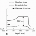
72 Gy (range 50 to 88 Gy) with a median fraction dose being 4.5 Gy (range 2.9 to 6 Gy). The proton dose at Tsukuba has been expressed in Gy as the uncorrected physical dose rather than in the relative biological effectiveness (RBE)-corrected Gy (RBE) (generally using an RBE of 1.1) which most proton facilities have employed. Patients were treated with different fractionation regimens. For that reason, equivalent doses at 2 Gy per fraction for the several fractionation regimens employed were calculated using a linear quadratic (LQ) model with α/β ratios of 10 and 3 for early and late responding tissues (see Table 19.4).9


TABLE 19.1 CARBON ION RADIOTHERAPY FOR GASTROINTESTINAL MALIGNANCIES AT THE NATIONAL INSTITUTE OF RADIOLOGICAL SCIENCES (NIRS) (APRIL 1995 TO FEBRUARY 2006)


Disease


Pathology/Diagnosis


Nature of Treatment


Number of Lesions


Hepatocellular carcinoma


Hepatocellular carcinoma


Carbon ion radiotherapy alone


200


Pancreatic cancer (operable)


Infiltrating pancreatic duct cancer


Resection after preoperative irradiation


33


64


Pancreatic cancer (inoperable)


Infiltrating pancreatic duct cancer


Carbon ion radiotherapy alone


31


Esophageal cancer


Squamous cell carcinoma of thoracic esophagus


Resection after preoperative irradiation


41


Postoperative, pelvic recurrence of rectal cancer


Postoperative, pelvic recurrence of rectal cancer


Carbon ion radiotherapy alone


67


Total number


372 (14.1%)


TABLE 19.2 CLINICAL CHARACTERISTICS OF 162 PATIENTS WITH HEPATOCELLULAR CARCINOMA TREATED WITH PROTONS AT TSUKUBA


Characteristics


Number of Patients (%)


Age (years)


<60


56 (34.6)


60-69


72 (44.4)


≥70


34 (21.0)


Gender


Men


124 (76.5)


Women


38 (23.5)


Underlying liver disorder


Cirrhosis


154 (95.1)


Class A


82 (50.6)


Class B


62 (38.3)


Class C


10 (6.2)


Chronic hepatitis


7 (4.3)


Normal liver


1 (0.6)


Etiology of liver disorder


Hepatitis B virus


15 (9.3)


Hepatitis C virus


129 (79.6)


Hepatitis B and C virus


3 (1.9)


Non-B, Non-C hepatitis


11 (6.8)


Alcoholic


2 (1.2)


Unknown


2 (1.2)


Performance status


0


61 (37.7)


1


79 (48.8)


2


21 (13.0)


3


1 (0.6)


4


0(0)


TABLE 19.3 CHARACTERISTICS OF THE HEPATIC TUMORS IN PATIENTS TREATED WITH PROTONS AT TSUKUBA


Characteristics


Number (%)


Number of tumors


Single


80 (49.4)


Multiple


82 (51.6)


Type of tumor


Nodular


156 (96.3)


Massive


6 (3.7)


Diffuse


0


TNM stage


Stage I


66 (40.7)


Stage II


70 (43.2)


Stage IIIA


25 (15.4)


Stage IIIB


1 (0.6)


Tumor size (cm)a


<3.0


51 (26.6)


3.1-5


108 (56.3)


>5.0


33 (17.2)


Serum α-fetoprotein (ng/mL)


<20


50 (30.9)


20-99


38 (23.5)


100-500


35 (21.6)


>500


39 (24.1)


a One hundred ninety two tumors treated using proton beam therapy.


TABLE 19.4 DOSE FRACTIONATION AND EQUIVALENT DOSES IF GIVEN AT 2 GY PER FRACTION IN PATIENTS TREATED WITH PROTONS AT TSUKUBA UNIVERSITY


Equivalent
Total Dose
(2 Gy/Fraction)


Total
Dose (Gy)


Number of
Fractions


Dose/Fraction


α/β
= 10


α/β
= 3


72


16


4.5


87.0


108.0


78


20


3.9


90.4


107.6


84


24


3.5


94.5


109.2


50


10


5.0


62.5


80.0


TABLE 19.5 RESULTS OF THE COX PROPORTIONAL HAZARD REGRESSION FOR DIFFERENT VARIABLES ON THE SURVIVAL OF PATIENTS TREATED WITH PROTONS AT TSUKUBA


Variable


Adjusted Rate Ratio


95% CIa of Hazard Ratio


p-Value


Child-Pugh Class


Class A


1.00


Class Bb


2.22


1.54-3.19


<0.0001


Class Cc


3.57


1.8-7.03


0.0002


Number of tumors


Solitary


1.00


Multiple


1.58


1.09-2.31


0.02


Tumor size (mm)


<50


1.00


≥50


1.41


0.92-2.15


0.11


Proton doses


<72 Gy


1.00


>72 Gy


1.01


0.74-1.46


0.95


Prior treatment


Received


1.00


Not received


0.88


0.58-1.34


0.56


a CI, confidence intervals.

b Values of Child-Pugh B compared with those of Child-Pugh A.

c Values of Child-Pugh C compared with those of Child-Pugh A.


The local control rate calculated using the Kaplan-Meier method at 5 years was 86.9% for all 192 tumors among the 162 patients. The overall survival rate for all 162 patients at 5 years was 23.5%. The degree of impairment in hepatic function attributable to coexisting cirrhosis and the number of tumors affected survival in the Cox regression analysis (see Table 19.5). Fifty patients had favorable prognostic features (Child-Pugh class A and a solitary tumor); their 5-year survival rate was 53.5%.

Treatment was generally well tolerated. The performance status (PS) of two (1.4%) patients declined from 0 before irradiation to 1 after irradiation and in another two patients (1.4%) from PS 2 before to PS 3 after irradiation. All remaining patients maintained an equivalent PS before and after irradiation. Table 19.6 shows that very few acute reactions to the treatment were seen among the 162 patients over 185 treatment courses, aside from the transient elevation of aspartate aminotransferase (AST) and alanine aminotransferase (ALT). All acute toxicity resolved within 2 weeks and no patients discontinued treatment because of acute side effects.

Five patients had late sequelae of grade II or higher (the late radiation morbidity scoring scheme of the Radiation Therapy Oncology Group/European Organization for Research and Treatment of Cancer [RTOG/EORTC]). One patient developed fibrotic stenosis of the common bile duct within the treatment volume, noted 13 months after irradiation. Two patients developed biloma with
infection adjacent to the irradiated volume at 29 and 38 months after irradiation, respectively. One patient developed an intractable gastric ulcer in the treatment volume 4 months after irradiation and another patient had an ulcer in the ascending colon 6 months after irradiation. No patients died of treatment sequelae in this series.


TABLE 19.6 TREATMENT SEQUELAE IN 162 PATIENTS GIVEN 185 COURSES OF PROTON THERAPY FOR HEPATOCELLULAR CARCINOMA AT TSUKUBA


Treatment Sequelae


Number of Treatment Courses (%)


Acute-subacute


Elevation of bilirubin


3 (2.1)


Anemia


2 (1.1)


Neutropenia


1 (0.5)


Thrombocytopenia


6 (3.2)


Elevated transaminase level


18 (9.7)


Late


Infectious biloma


2 (1.1)


Common bile duct stenosis


1 (0.5)


Gastrointestinal tract bleeding


2 (1.1)


Figure 19.1 Using percutaneous ultrasound-guided biopsy, a 26-year-old man was diagnosed with a hepatocellular carcinoma in the right lobe of the liver. He underwent transcatheter arterial infusion of chemotherapy with lipiodol in October 2001 and segmental hepatectomy of the right lobe in November 2001. He was treated with proton beam therapy after the second course of transcatheter arterial infusion of chemotherapy with lipiodol for a recurrent tumor in December 2002. He received a total proton dose of 60 Gy in 10 fractions over 2 weeks to the tumor in the S3 segment of the left lobe through anterior and left anterior oblique ports (arrows in A) in 2003. The tumor was controlled locally for more than 3 years by 2006. Note that the volume of the unirradiated portion of the liver enlarged (C). The dose distribution generated using two proton beams through the anterior and the left anterior oblique ports in the axial dimension is shown in B. Isodose distribution curves in red and in blue respectively represent 100% and 10% of the total dose.

From September 2001 through August 2004, the new facility treated 153 patients with HCC. Of those patients, 52 were treated with a proton dose of 60 Gy in 10 fractions over 2 weeks. The patients had Child-Pugh class A or B liver function and a solitary tumor of 10 cm or less in maximal diameter at the peripheral region of the liver. Of the 52 patients, 34 were men and 18 women; their median age was 69 years (range 26 to 85 years); 39 patients had Child-Pugh class A and 13 had B. The median tumor maximal diameter was 2.8 cm (range 0.8 to 9.3 cm). The median observed period was 22 months (1 to 47 months). Thirty-two patients were alive as of March 2006; 20 had died. The 3-year survival and local control rates were 41.9% and 94.6%, respectively. Grade 2 or higher late complications were observed in two patients—one patient suffered from radiation pneumonitis (grade 2) and rib fracture at 1 and 27 months after PBT; the other suffered from rib fracture at 8 months after PBT. This irradiation regimen of 60 Gy in 10 fractions is safe and effective when the tumor is located in the peripheral region of the liver and the liver function is reasonably preserved.

Clinical images for a representative case are shown in Figures 19.1A and 19.1B.


Repeated Proton Beam Therapy

Multifocal carcinogenesis characterizes HCC in a cirrhotic liver. For that reason, patients frequently undergo repeated treatments during the course of an illness.8,10 More than 80% of patients who undergo surgery or PBT eventually develop another HCC within 5 years.8,11

Only gold members can continue reading. Log In or Register to continue

Stay updated, free articles. Join our Telegram channel

Aug 27, 2016 | Posted by in GENERAL RADIOLOGY | Comments Off on Gastrointestinal Malignancies

Full access? Get Clinical Tree

Get Clinical Tree app for offline access