Fig. 28.1
Radiographic assessment of disproportional skeletal growth. (a) Spine. (b) Extremities
28.4.2 Achondroplasia
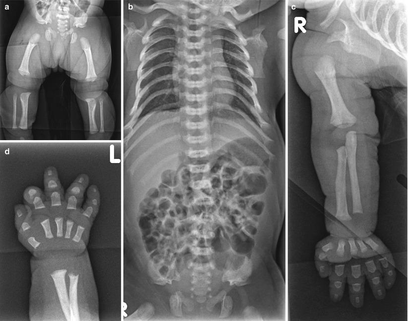
Fig. 28.2
Achondroplasia: newborn. (a) Anteroposterior view of the pelvis and lower extremities shows horizontal acetabula, square iliac bones, and short sacrosciatic notches. Note the oval transradiancy of proximal femora and sloping metaphyses showing ice cream scoop appearance. (b) Anteroposterior view of the spine shows slightly flat vertebral bodies and increased height of the intervertebral space. The interpediculate distance decreases from the upper to the lower lumbar spine. (c) Rhizomelic shortening is evident in the upper extremity. (d) Hand radiograph demonstrates wide separation of the third and fourth phalanges suggesting a trident hand
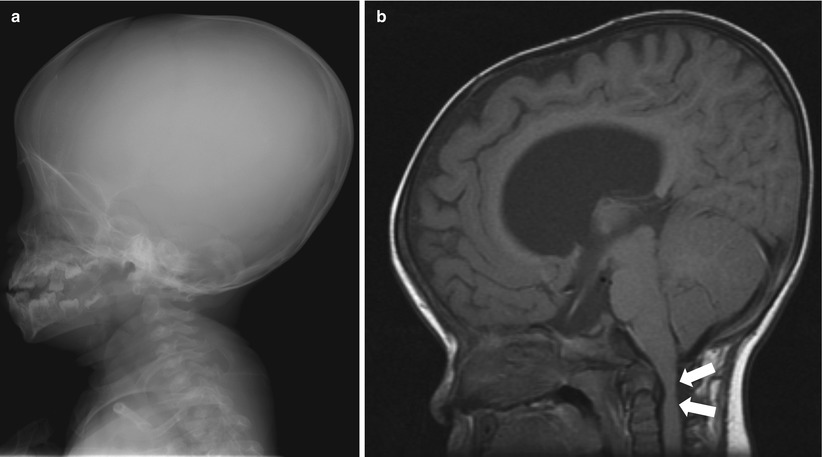
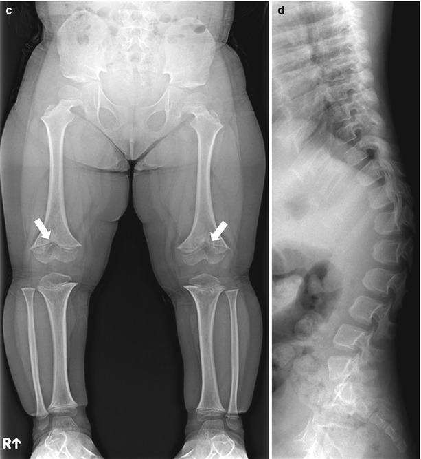
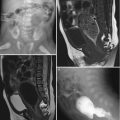
Fig. 28.3
Achondroplasia: older child. (a) Skull lateral view of a 3-year-old boy shows macrocephaly and narrow skull base with J-shaped sella. Note the frontal bowing and midface hypoplasia. (b) Sagittal T1-weighted image of the same patient demonstrates hydrocephalus and narrow formen magnum (arrows). (c) Lower extremity radiograph of a 6-year-old boy shows short tubular bones and mild metaphyseal flaring with V-shaped distal femoral growth plate (arrow). (d) Lateral view of the spine in a 4-year-old boy shows variable degrees of anterior wedging of the vertebral bodies, causing gibbus. The posterior aspect of the vertebral bodies is concave, and the pedicles are short
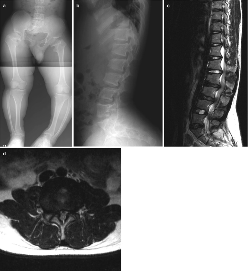
Fig. 28.4
Achondroplasia: adolescent. (a) Pelvis and lower extremity radiograph of a 17-year-old boy shows typical champagne glass–type pelvic inlet with squared iliac wing and horizontal acetabular roof. Short and thick tubular bones, short femoral neck, and left genu varum deformities are seen in the lower extremities. (b) The lateral view of thoracolumbar spine in a 13-year-old girl shows posterior and scalloping of the vertebral bodies and narrow spinal canal. There is increased angulation at the lumbosacral junction. (c–d) T2-weighted sagittal (c) and axial images (d) of the same patient show diffuse narrowing of the spinal canal at the level of lumbar spine and posterior scalloping of the vertebrae
28.4.3 Spondyloepiphyseal Dysplasia Congenita
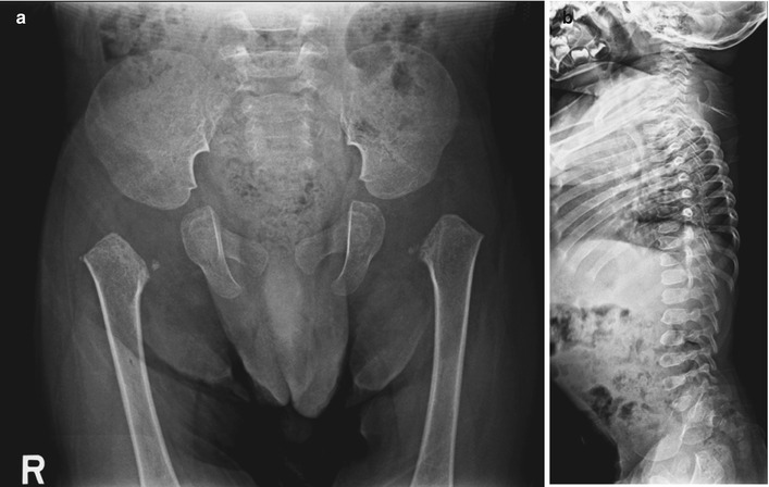
Fig. 28.5
Spondyloepiphyseal dysplasia congenita. (a) Anteroposterior view of pelvis in a 20-month-old boy shows short pubic rami and small femoral secondary ossification center. (b) Lateral view shows flat vertebral bodies. The thoracolumbar vertebral bodies show dorsal wedging giving them a “pear-shaped” appearance
28.4.4 Kniest Dysplasia
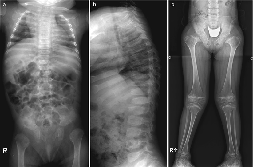
Fig. 28.6
Kniest dysplasia. (a–b) Spine radiographs of an 8-month-old girl show flat vertebrae with anterior wedging. The ilia are short and proximal epiphyses are not ossified. (c) Anteroposterior view of a 7-year-old girl shows broad and short femoral neck and delayed femoral head ossification. The epiphyses at the knee are large, and the growth plate of the tibiae has an inverted V shape
28.4.5 Metatropic Dysplasia
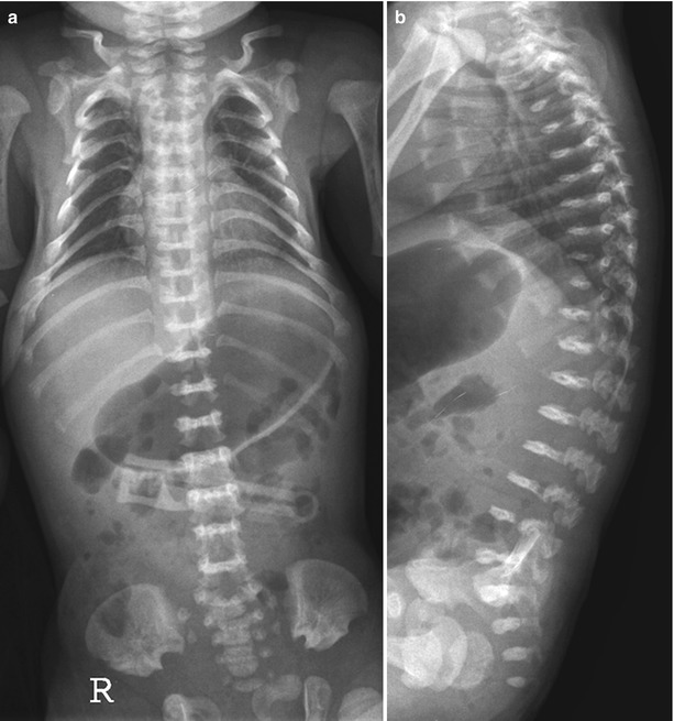
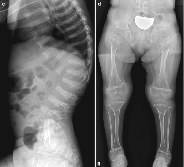
Fig. 28.7
Metatropic dysplasia. (a–b) Newborn. Anteroposterior and lateral views of thoracolumbar spine show marked platyspondyly giving “wafer-thin vertebra”. (c) Four years. Lateral view of thoracolumbar spine shows exaggerated thoracolumbar kyphosis and platyspondyly. (d) The iliac wings are crescent shaped, the lower portions of the ilia are hypoplastic, and sacrosciatic notches are narrow. The acetabular roofs are broad and horizontal and the pubic and ischial bones, short and thick. The tubular bones are short and flared at both ends giving “dumbbell-shaped” femora
28.4.6 Short Rib Polydactyly
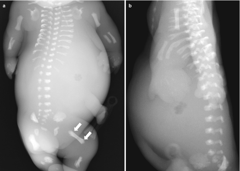
Fig. 28.8
Short rib polydactyly. (a) Stillbirth. Anteroposterior view of the chest and abdomen shows horizontally oriented, severely shortened ribs with restricted thoracic cage and protruded abdomen. The long tubular bones are shortened with spurs of bone extending longitudinally, giving a “banana peel” appearance (arrows). (b) Lateral view of the spine shows small vertebral bodies with irregular end plates
28.4.7 Asphyxiating Thoracic Dysplasia
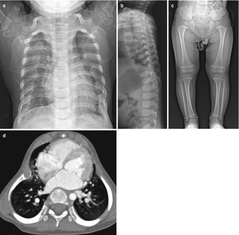
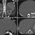
Fig. 28.9
Asphyxiating thoracic dysplasia. (a–b) 1 year. Anteroposterior view of chest and spine lateral view show moderately short ribs with irregular costochondral junctions, narrow thorax, and handle bar appearance of the clavicles. (c) The iliac bones are short in cephalocaudal dimension. There are metaphyseal irregularities in the tubular bones. (d) Chest CT demonstrates deformed thoracic cage with small lung volume
28.4.8 Pseudoachondroplasia
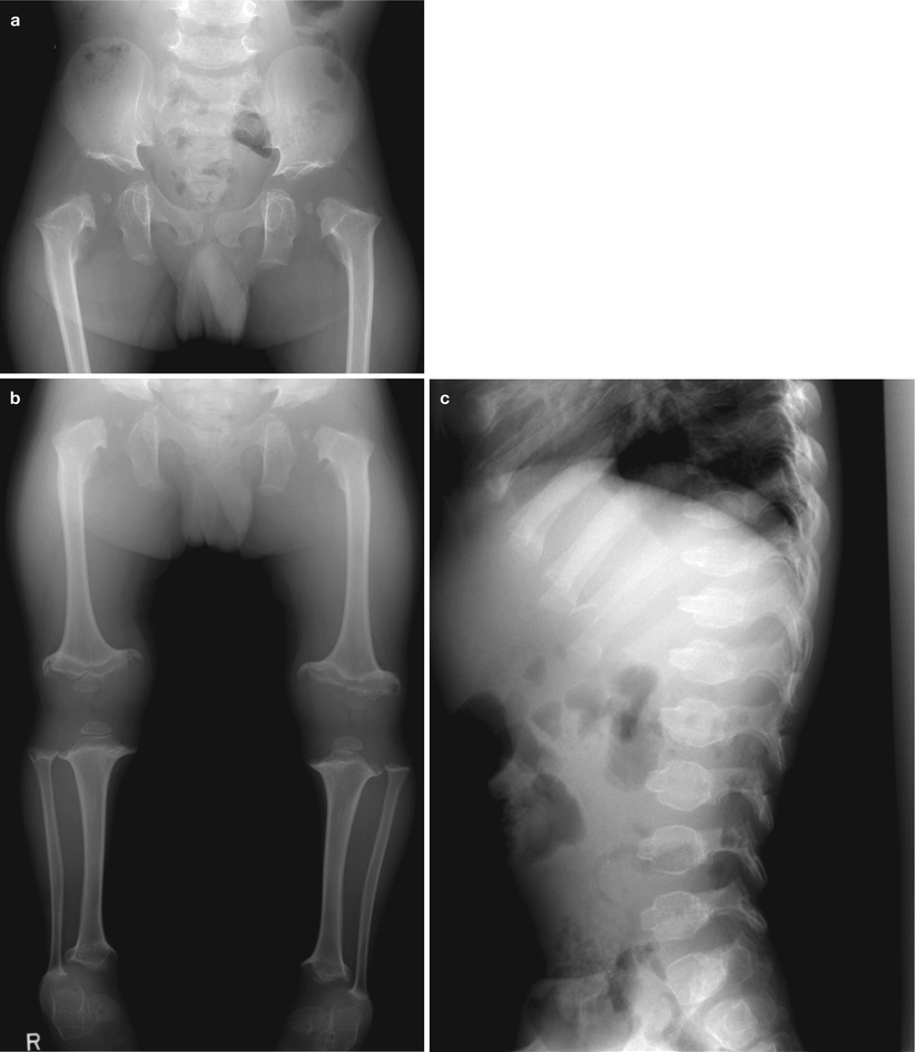
Fig. 28.10
Pseudoachondroplasia. (a) Anteroposterior view of pelvis shows deep acetabula, long femoral neck, and small femoral head ossification center. (b) The tubular bones are severely shortened with wide, irregular metaphyses. (c) Lateral view of the thoracolumbar spine shows an anterior protrusion of the central aspects of the vertebral bodies and a biconvex configuration of the upper and lower vertebral plates
28.4.9 Multiple Epiphyseal Dysplasia
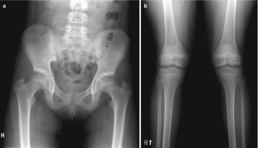
Fig. 28.11
Multiple epiphyseal dysplasia. (a–b) Anteroposterior views show small proximal femoral head ossification and flattened and irregular distal femoral epiphysis
28.4.10 Metaphyseal Chondrodysplasia
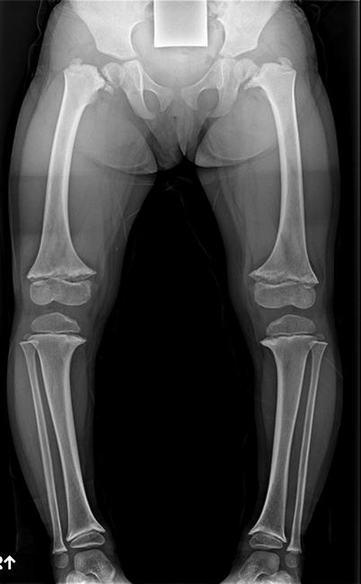
Fig. 28.12
Metaphyseal chondrodysplasia. A 3-year-old girl. The femoral necks are short and in various positions. The physeal plates are wide and irregular
28.4.11 Shwachman-Diamond Syndrome
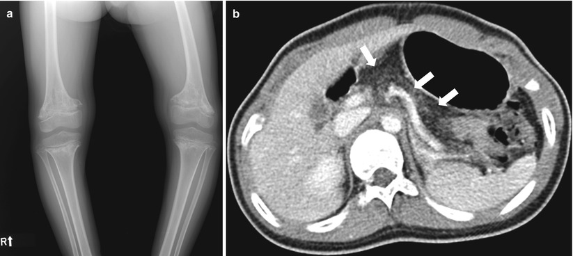
Fig. 28.13


Shwachman-Diamond syndrome. (a) Metaphyseal ossification is markedly irregular with splaying and fraying of the metaphyses. (b) Abdomen CT demonstrates pancreatic lipomatosis (arrows)
Related posts:


Stay updated, free articles. Join our Telegram channel
Full access? Get Clinical Tree
