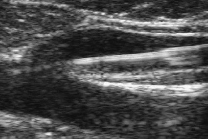
Great Saphenous Vein Ablation
Robert J. Min and Neil M. Khilnani
Chronic venous insufficiency (CVI) is extraordinarily common, with estimates of up to 25% of women and 10% of men suffering from some form of CVI.1 Most patients with CVI have symptoms that interfere with daily living (e.g., leg aches, fatigue, throbbing, heaviness, night cramps). Severe cases can lead to skin damage resulting from chronic venous hypertension (e.g., eczema, edema, hyperpigmentation, lipodermatosclerosis).
The majority of patients with leg ulceration have superficial venous insufficiency (SVI) as the primary underlying cause, with SVI being the sole factor in 20%.2
Patients with symptoms typical of CVI and clinical signs of CVI require further evaluation with duplex ultrasound (DUS).3,4 The goal of DUS evaluation is to map out all the incompetent venous pathways responsible for the patient’s condition, including the primary or highest points of reflux and the presence of obstruction.3 Such a map is necessary to determine the best treatment plan.
Dr. Boné first reported on delivery of endoluminal laser energy in 1999.5 Since then, a method for treating the entire incompetent vein segment has been described by Min and Navarro.6–8 Endovenous laser treatment, which received approval by the U.S. Food and Drug Administration (FDA) in January 2002, achieves nonthrombotic vein occlusion by delivery of laser energy directly into vein walls. Lasers with wavelengths of 810, 940, 980, 1064, and 1320 nm have all been used with success. Contact between the laser fiber and vein wall is necessary to cause sufficient damage to the vein to result in acute wall thickening with eventual vein contraction and fibrosis.
Indications
Medical treatment should be contemplated when varicose vein symptoms persist despite conservative methods such as graduated compression and exercise. Other indications may include treatment or prevention of complications arising from chronic venous hypertension, such as bleeding, superficial thrombophlebitis, and skin damage. Patients who are unable or unwilling to tolerate long-term compression or those desiring cosmetic improvement may also be candidates for medical intervention. Indications for endovenous ablation of incompetent truncal veins are similar to those for surgical ligation and stripping.
Contraindications
Many of the exclusion criteria for sclerotherapy are also relative contraindications to endovenous treatment, including dependency on the saphenous system for venous drainage because of significant deep venous obstruction, active deep venous thrombosis (DVT), nonpalpable pedal pulses, inability to ambulate, general poor health, or women who are pregnant or nursing. Additional relative contraindications to all catheter-based endovenous ablation techniques are nontraversable vein segments as a result of either thrombosis or extreme tortuosity. Fortunately, these findings are uncommon and should be recognized on pretreatment venous DUS mapping.
Equipment
The equipment necessary to perform endovenous laser ablation includes but is not limited to:
Technique
Anatomy and Approach
Before treatment, the abnormal venous pathways are mapped with ultrasound guidance, and the veins to be treated are marked on the overlying skin with the patient standing. Additional important landmarks such as the entry point, junctions, aneurysmal segments, and areas of significant blood flow from tributaries or perforators should be noted.
Endovenous ablation of the great saphenous vein (GSV) is performed with the patient in the supine or oblique position and the hip slightly turned to expose the course of the GSV. When the small saphenous vein (SSV) is the target, patients are placed prone with their feet hanging off the end of the table to relax the calf muscle and popliteal fossa. Treating multiple sources of venous reflux may require multiple repositioning and repeat preparation.
In almost all cases, the target vein is entered or access is gained via one of its direct tributaries. Entry through a tributary should be attempted only if the vein is relatively straight and of sufficient diameter, because these veins tend to be more prone to venospasm and can be more difficult to traverse. In general, the vein is punctured at or just peripheral to the lowest level of truncal reflux as determined by DUS. At this point, saphenous vein diameter abruptly decreases, and it regains its competence after escape of the refluxing blood through incompetent varicose tributaries. Saphenous vein incompetence can occur segmentally. Reflux escaping through a tributary vein can reenter the saphenous vein at a lower level. In this case, the lowest incompetent vein segment should be accessed and all refluxing vein segments ablated via one puncture. At times, however, more than one puncture may be necessary.
Technical Aspects
The target vein is entered with either a 19- or 21-gauge needle under real-time ultrasound guidance with a single-wall technique. Using reverse Trendelenburg positioning and keeping the procedure room warm until access is achieved will help minimize shrinkage. Other ancillary procedures advocated by some practitioners to maximize vein size include a heating pad or a small amount of nitroglycerin paste at the access point. Non-GSV segments are especially prone to venospasm, so particular care must be taken when accessing a tributary vein segment or a truncal vein such as the anterior accessory GSV, SSV, or thigh circumflex vein.
A 5F vascular introducer sheath with markings is inserted over a guidewire into the vein and passed through the entire abnormal segment into a more central vein. A bare-tipped laser fiber is inserted into the sheath. The sheath is then pulled back to expose the tip of the fiber, and the fiber is locked in place. Under ultrasound guidance, the introducer sheath and fiber are withdrawn out of the deep veins and positioned within the superficial venous system at the junction, as seen in Figure 106-1. The fiber is left in this position during tumescent anesthetic administration and will be repositioned just before delivery of laser energy. Confirmation of position can be made by direct visualization of the red aiming beam through the skin.

An important part of the procedure is correct delivery of perivenous tumescent anesthesia. Proper use of tumescent anesthesia should make endovenous laser therapy painless, without the need for intravenous sedation or general anesthesia. In fact, it can be argued that sedation adds risk to endovenous laser ablation by blunting patient feedback during the procedure, as well as delaying immediate postprocedure ambulation.
In addition to making the procedure painless, tumescent anesthesia is used to maximize the safety and efficacy of endovenous laser treatment (EVLT). Although venospasm may occur in some veins, proper delivery of tumescent fluid into the perivenous space will ensure compression of the vein around the laser fiber and achieve contact between the vein walls and laser fiber tip. This will allow adequate transfer of laser energy to the target vein walls and result in vein wall damage and subsequent fibrosis. Inadequate vein emptying with too much blood remaining within the vein will lead to suboptimal heating of the vein wall. In this case, the occlusion may be due to thrombosis, which will inevitably result in recanalization.
Many practitioners believe the surrounding cuff of tumescent fluid also serves as a protective barrier to prevent heating of nontarget tissues, including skin, nerves, arteries, and deep veins. Injection of tumescent fluid in the proper plane can be achieved only with DUS guidance. The tumescent fluid should be delivered between the target vein and adjacent nontarget tissues. We prefer to deliver the tumescent anesthetic by hand pressure from a 25-gauge needle attached to a 20-mL syringe. For right-handed operators, the tumescent fluid is injected distally to proximally. Skin punctures are required every 3 to 5 cm until the proper perivenous tissue plane is located. Once this occurs, fluid will track more easily up and around the target vein, and greater distances can be covered with each needle puncture.
To treat a 45-cm segment of vein, approximately 100 to 150 mL of 0.1% lidocaine neutralized with sodium bicarbonate may be required. This mixture can be made by diluting 50 mL of 1% lidocaine in 450 mL of normal saline and adding 5 to 10 mL of 8.4% sodium bicarbonate. If it is anticipated that larger volumes of tumescent anesthetic will be necessary, a concentration of 0.05% lidocaine can be used effectively. These amounts of lidocaine are well within the safe doses of 4.5 mg/kg of lidocaine without epinephrine and 7 mg/kg with epinephrine. Although many practitioners choose to use lidocaine with epinephrine to maximize venospasm and minimize bruising, we achieve adequate and complete vein emptying with plain lidocaine and avoid the risk of toxicity related to epinephrine.
After administration of tumescent anesthesia, ultrasound is used to check for adequacy. A centimeter halo of fluid surrounding the target vein or separating the vein from the overlying skin has been found to be sufficient, as demonstrated in Figure 106-2. Proper delivery of tumescent fluid may be particularly useful for maximizing procedural safety when performing endovenous laser therapy in certain locations such as tributaries close to the skin, the SSV near the saphenopopliteal junction, or the GSV below the knee, owing to the close proximity of nerves or arterial branches. Adequacy of separation of arterial branches from the target vein can be checked with color Doppler ultrasound.

Stay updated, free articles. Join our Telegram channel

Full access? Get Clinical Tree

