Hallux Valgus Deformity
Anatomic Considerations
The metatarsophalangeal joints are condyloid joints characterized by the articulation of the rounded articular surfaces of the metatarsal heads into the shallow concavities of the articular surfaces of the proximal end of the first phalanges (Fig. 19.1). Each joint is lined with synovium and the ample synovial space allows for intra-articular placement of needles for injection and aspiration. The metatarsophalangeal joints have a dense joint capsule and strong plantar and collateral ligaments, although fracture and subluxation may still occur (Figs. 19.2 and 19.3). The metatarsophalangeal joints are also susceptible to overuse and misuse injuries with resultant inflammation and arthritis (Fig. 19.4).
Clinical Correlates
Bunion, which is also known as the hallux valgus, refers to a constellation of symptoms including soft tissue swelling over the first metatarsophalangeal joint associated with abnormal angulation of the joint that results in a prominent first metatarsal head (Fig. 19.5). Ultimately, the first metatarsophalangeal joint may sublux, and the overlapping of the first and second toes worsens, resulting in a painful condition known as the hallux valgus deformity (Fig. 19.6). The metatarsocuneiform joint may also play a role in the evolution of hallux valgus (Fig. 19.7). Frequently an inflamed adventitious bursa may coexist with the bunion, further exacerbating the pain and cosmetic deformity (Fig. 19.8). More common in women, bunion is usually the result of wearing shoes with a too-tight toe box, with the wearing of high-heeled shoes exacerbating the problem.
 FIGURE 19.1 Anatomy of the metatarsophalangeal joints. |
Most patients who present with bunion also present with the complaint of pain that is localized to the affected first metatarsophalangeal joint and the inability to get shoes to fit. Walking makes the pain worse, with rest and heat providing some relief. The pain is constant and is characterized as aching and may interfere with sleep. Some patients complain of a grating or popping sensation with use of the joint, and crepitus may be present on physical examination. In addition to the just-mentioned pain, patients who suffer with bunions develop the characteristic hallux valgus deformity, which consists of a prominent first metatarsal head and improper angulation of the joint, with overlapping first and second toes.
Functional disability often accompanies the pain of the hallux valgus joint. Patients will often notice increasing difficulty in performing their activities of daily living and tasks that require standing, walking, or weight bearing. If the pathologic process responsible for pain of hallux valgus is not adequately treated, the patient’s functional disability may worsen and muscle wasting and ultimately a frozen first metatarsophalangeal joint may occur.
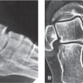
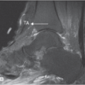
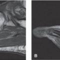
Plain radiographs are indicated in all patients who present with pain of the hallux valgus (Fig. 19.9). Based on the patient’s clinical presentation, additional testing may be indicated including complete blood cell count, sedimentation rate, and antinuclear antibody testing. MRI or ultrasound of the hallux valgus joint is indicated if fracture, effusion, tendinopathy, crystal arthropathy, joint mice, synovitis, foreign body, bursitis, or ligamentous injury is suspected (Fig. 19.10).
Related posts:
 Arthritis and Other Abnormalities of the Subtalar Joint
 Arthritis and Other Abnormalities of the Ankle Joint
 Anterior Tarsal Tunnel Syndrome and Other Abnormalities of the Deep Peroneal Nerve
 Retrocalcaneal and Achilles Bursitis
 Calcaneal Spurs
 Plantar Fasciitis and Other Abnormalities of the Plantar Fascia
Stay updated, free articles. Join our Telegram channel

Full access? Get Clinical Tree


