Hands and Feet: Osseous Alterations


10.1055/b-0034-87939

Hands and Feet: Osseous Alterations


Table 5.60 Hands and feet: alterations in size, shape, contour, or architecture

Diagnosis


Findings


Comments


Osteosclerosis


Fig. 5.101


Increased focal density with a narrow zone of transition.


DD: enostosis (bone island), terminal phalangeal sclerosis (associated with JIA), melorheostosis.


Enchondroma


Fig. 5.102


Fig. 5.103, p. 564


(see Table 5.71 )


Periostitis


(see Table 5.81 )


Elongation, megadactyly


(see Table 5.95 )


Slender phalanges, arachnodactyly


Long and slender fingers.


Homocystinuria, Marfan syndrome, congenital arachnodactyly with contractures.

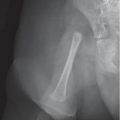
Fig. 5.101 Phalangeal osteosclerosis (differental diagnosis includes enostosis and melorrheostosis).
Fig. 5.102 Enchondroma in the proximal phalanx of the fourth toe (arrow).
Fig. 5.103 Enchondroma of the proximal phalanx of the toe with a pathologic fracture.


Table 5.61 Hands and feet: alterations in distal phalanges

Diagnosis


Findings


Comments


Clubbing


Bulbous thickening of the ends of the fingers. Confined to the soft tissues and spares the bones.


Cyanotic heart disease, chronic lung disease, lung tumors, pachydermoperiostosis, chronic gastrointestinal disease, gigantism.


Hyperplasia


Widening of the tuft with a narrow shaft (drumstick appearance).


Normal variant, Coffin-Lawry (drumstick terminal phalanges), Holt-Oram syndrome (triphalangeal thumb).


Hypoplasia


(see Table 5.59 )


Acroosteolysis


Fig. 5.104


Resorption or erosions of the distal phalanges, especially the tufts.


Congenital: brachydactyly type B (see Table 5.59 ). DD: trauma, infection, frostbite, leprosy, psoriasis, hyperuricemia, polyvinyl toxicity, insensitivity to pain, epidermolysis bullosa, secondary hyperparathyroidism.

Fig. 5.104 Acroosteolysis. A 3-year-old with resorptive metabolic bone disease of unclear etiology.


Table 5.62 Hands and feet: digit I

Diagnosis


Findings


Comments


Brachydactyly isolated


(see Table 5.59 )


Radial ray anomalies


Fig. 5.86, p. 553


VACTERL, Holt-Oram syndrome, thrombocytopenia absent radius syndrome, Fanconi pancytopenia.


Triphalangeal thumb


Thumb has an extra phalanx. DD: Blackfan-Diamond syndrome, Holt-Oram syndrome, trisomy


13, Poland syndrome, VATER, Werner mesomelic dysplasia, Juberg-Hayward syndrome, thalidomide and phenytoin embryopathy, and sporadic (rarely). Associated with duplication or absence of the contralateral thumb.


Acrocephalosyndactyly (Apert syndrome)


Fig. 5.105, p. 566


Widened, short thumbs. Thumbs and great toes may also be duplicated.


Craniosynostosis, midface hypoplasia, and syndactyly of the hands and feet. Crouzon and Pfeiffer syndromes are allelic disorders with overlapping features.


Rubinstein-Taybi syndrome


Widened, short thumbs. Thumbs and great toes may also be duplicated.


Characterized by mental retardation, broad thumbs and toes, and facial abnormalities.


Other syndromes


Abnormal shape (usually short or broad) thumbs and great toes.


Nonspecific finding seen in otopalatodigital syndrome, frontodigital syndrome, hand-foot-uterus syndrome, progressive myositis ossifi-cans, diastrophic dysplasia (hitchhiker thumb), Larsen syndrome, Leri pleonosteosis, hemifacial microsomia (Goldenhar syndrome), trisomy 13 (polydactyly), Poland syndrome (syndactyly), thalidomide embryopathy, Juberg-Hayward syndrome, etc.

Fig. 5.105 Apert syndrome with syndactyly, fusion of metacarpals, and hypoplasia of the thumb.


Table 5.63 Hands and feet: digit V

Diagnosis


Findings


Comments


Kirner (dystelephalangy)


Radial bowing distal phalanx of the fifth finger. Usually bilateral.


Differentiate from camptodactyly (flexure contraction) and type A3 brachydactyly.


Clinodactyly


Radial bowing of the fifth finger.


Most often a sporadic incidental finding. DD: includes genetic syndromes such as trisomy 21, Russell-Silver, Feingold, Robinow, and Cornelia de Lange syndromes (see Table 5.59 and brachydactyly type A3).


Senior syndrome


Shortening of middle phalanx of the fifth finger, fusion of middle and distal phalanges of the fifth toe, and absence of middle and distal phalanx of fifth toe.


Short children with tiny toenails.


Coffin-Siris syndrome


Absence of the distal phalanges of the fifth fingers and fifth toes.


Mental retardation with absent nail and terminal phalanx of the fifth finger.


Table 5.64 Hands and feet: metacarpals and metatarsals

Diagnosis


Findings


Comments


Idiopathic


Short fourth and fifth metatarsals.


Seen in 10% of normal population.


Radial ray anomalies


Hypoplastic thumb metacarpal.


The radial ray anomaly may be confined to the first metacarpal.


Turner syndrome


Short fourth and fifth and ± third metacarpals.


Sickle cell disease


Shortening secondary to infarction.


Pseudohypoparathyroidism, pseudopseudohypoparathyroidism


Short fourth and fifth metacarpals.


Subcutaneous calcifications. Shortening not seen in pseudohypoparathyroidism types 1b and 2.


Homocystinuria


Short fourth metacarpal.


Other disorders associated with short metacarpals or metatarsals


Over 200 syndromes are associated with short metacar-pals or metatarsals.


Table 5.65 Hands and feet: carpal and tarsal bones

Diagnosis


Findings


Comments


Coalition/fusion


Fibrous or osseous union between one or more carpal or tarsal bones.


Sporadic, acquired (trauma, JIA), or associated with syndromes (Holt-Oram, Turner, arthrogryposis, acrocephalosyndactyly).


Pseudocyst, intraosseous lipoma, bone cyst, osteonecrosis


Well-defined region of lucency in the distal calcaneus.


MRI may help with the differential diagnosis.


JIA, infection, pigmented villonodular synovitis


Fig. 5.106a–c


Multiple cystic lucencies.


Spotty carpus.

Fig. 5.106a–c Juvenile idiopathic arthritis and spotty carpus. T1- (a) and T2-weighted (b) MRI show osteitis and separate regions of synovium that have eroded into several carpal bones (arrows). (c) The intraosseous synovitis enhances with gadolinium (arrow).

Only gold members can continue reading. Log In or Register to continue

Stay updated, free articles. Join our Telegram channel

Jul 12, 2020 | Posted by in PEDIATRIC IMAGING | Comments Off on Hands and Feet: Osseous Alterations

Full access? Get Clinical Tree

Get Clinical Tree app for offline access