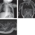
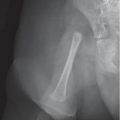
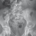
Fig. 5.107 Myositis ossificans in the vastus medialis after trauma (arrows).Fig. 5.108a–c Juxtacortical myositis ossificans. (a, b) One month after trauma, a region of extraskeletal ossification has developed adjacent to the proximal fibula (arrow in b). (c) Increased fluid signal intensity is seen between the new bone formation and underlying fibula on T2-weighted MRI (arrow).Fig. 5.109a–c Hemangioma with phleboliths (a) produces saucerization of the distal fibula. MRI shows avid enhancement and periosteal reaction on T1-weighted fat-suppressed imaging (b) after the administration of gadolinium and the contributions of blood vessels on time-resolved imaging with contrast kinetics (c).
Table 5.67 Hands and feet: soft-tissue calcifications (generalized)