SONOGRAM ABBREVIATIONS
Bl Bladder
Pr Prostate gland
KEY WORDS
Benign Prostatic Hypertrophy. Enlargement of the glandular component of the prostate. The true prostate forms a shell around the enlarged gland.
Cystitis. Infection or inflammation of the wall of the bladder.
Hematuria. Blood in the urine.
Hydroureter. Dilated ureter.
Nephrocalcinosis. Multiple small calculi deposited in the renal pyramids. Found in association with renal tubular acidosis and medullary sponge kidney.
Pyelonephritis. Infection of the kidney without abscess formation.
Pyonephrosis. Pus-filled hydronephrosis.
Staghorn Calculus. A stone that occupies most of the renal pelvis and infundibulum.
Trigone. Base of the bladder. Area between the insertion of the ureters and the urethra.
Urethra. Urinary outflow tract below the bladder.
RELEVANT LABORATORY VALUES
Microscopic hematuria: More than two to three red blood cells per high-power field prompts a full diagnostic workup, whereas one to three red blood cells per high-power field may warrant a workup in patients at risk for urologic malignancy.
The Clinical Problem
Hematuria is an important sign of genitourinary tract problems. The abnormality may be located within the kidneys, ureter, bladder, or urethra. The segments of the genitourinary tract that can be visualized by ultrasound (i.e., the kidney, upper and lower portions of the ureter, bladder, and upper part of the urethra) should be carefully examined. A specialized computed tomography (CT) scan (so-called CT-intravenous pyelogram [IVP]) is the gold standard, gradually replacing the older excretory urogram (IVP or intravenous urogram [IVU]), for evaluating patients with hematuria. Ultrasound, however, particularly when combined with a plain film, offers a safe and less-expensive method for evaluation of patients with suspected stone disease who cannot have contrast or who do not have access to modern CT. Good sonographic scanning technique is essential because the responsible lesions, such as calculi or tumors, are often relatively minute.
Anatomy
KIDNEYS
See Chapter 17.
BLADDER
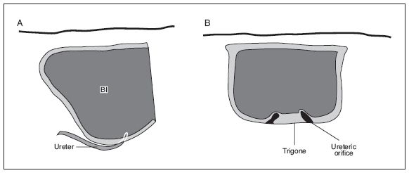
The bladder has a muscular wall whose thickness can be discerned with ultrasound. It is usually symmetric and is more or less square on transverse section. The base of the bladder where the ureters enter is known as the trigone (Fig. 20-1).
The bladder wall is proportionately thicker in infants than in other age groups. The bladder wall is approximately 3 mm thick when distended and 5 mm thick when empty in individuals other than infants.
The normal bladder in an adult contains approximately 150 to 400 mL and empties nearly completely.
URETERS
The ureters are the small tubes that convey urine from the kidney to the bladder. Each begins where the renal pelvis narrows (ureteropelvic junction) and travels anterior to the psoas muscle into the true pelvis. They insert into the trigone region of the bladder (ureterovesicular junction). The normal ureter measures less than 8 mm wide and is difficult to see with ultrasound under normal conditions. In hydroureter, the entire length of the ureter may be seen.
Two small bumps on the posterior aspect of the bladder on either side of the midline represent the ureteric orifices (see Fig. 18-1). The jet phenomenon may be seen when the ureteric orifices open and allow the emptying of urine from the ureters into the bladder. Color Doppler is a useful tool to demonstrate ureteral jets. The normal ureter can be seen as a subtle echopenic tube leading obliquely superiorly from the ureteric orifice. It distends with peristalsis.
URETHRA
The urethra is the tube that conveys urine from the bladder to the outside of the body. Ultrasound evaluation is generally limited to the posterior urethra above the external sphincter. In the male, the posterior urethra is surrounded by the prostate. The anterior urethra in the male can be scanned with a specialized technique in which aqueous gel is injected into the urethra and a linear transducer is placed parallel to the urethra on the penile shaft.

Figure 20-1. ![]() Ureteric orifices can be seen as two small buds in the trigone. Ureters can be traced through the wall to the ureteric orifice. A. Sagittal. B. Transverse.
Ureteric orifices can be seen as two small buds in the trigone. Ureters can be traced through the wall to the ureteric orifice. A. Sagittal. B. Transverse.
MALE ANATOMY
There are two major parts of the urethra, the posterior (most of which lies within the prostate gland) and the anterior (which courses through the penis). Posterior to the lower bladder lie the mustache-shaped seminal vesicles. The prostate lies posterior to the symphysis pubis and inferior to the bladder and seminal vesicles. It is more or less round. Sometimes a line of echoes from the urethra can be seen at its center.
FEMALE ANATOMY
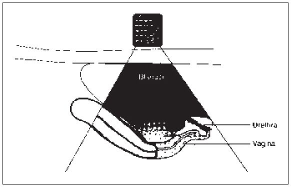
In the female, the uterus and vagina lie posterior and inferior to the bladder (Fig. 20-2). The vagina and lower segment of the uterus are normally never separated from the bladder. A tissue similar to the prostate in the male lies around the female urethra (Fig. 20-2) and can occasionally be mistaken for a mass in the base of the bladder.

Figure 20-2. ![]() In the female, the uterus and vagina lie posterior and inferior to the bladder. The posterior urethra is seen as a mass at the inferior aspect of the bladder.
In the female, the uterus and vagina lie posterior and inferior to the bladder. The posterior urethra is seen as a mass at the inferior aspect of the bladder.
Technique
KIDNEY
The sonographic examination of the kidney is reviewed in Chapter 18.
BLADDER
Stay updated, free articles. Join our Telegram channel

Full access? Get Clinical Tree


