KEY FACTS
Terminology
- •
Hepatocellular adenoma (HCA) or liver cell adenoma
- •
2nd most frequent hepatic tumor in young women after focal nodular hyperplasia (FNH)
Imaging
- •
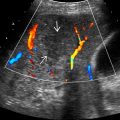
Heterogeneous, hypervascular, hypo-/iso-/hyperechoic mass
- •
Round or mildly lobulated, well-defined borders
- •
Subcapsular right hepatic lobe (75%)
- •
Complex heterogeneous mixed echogenicity mass
- •
Hypoechoic halo of compressed liver tissue with multiple vessels
- •
Larger lesions prone to intratumoral hemorrhage or rupture with intraperitoneal hemorrhage
- •
Best imaging tool: Ultrasound for lesion detection, guiding biopsy, and monitoring size
- •
Liver-specific MR contrast agents have high accuracy in distinguishing HCA from other liver lesions
Top Differential Diagnoses
- •
Hemangioma
- •
FNH
- •
Hepatocellular carcinoma
Clinical Issues
- •
Potential complications: ± hemorrhage, malignant degeneration
- •
More common in younger women on oral contraceptive pill (OCP)
- •
Definitive diagnosis and subtype with biopsy
- •
Molecular classification (4 subtypes) helps determine prognosis and management
- ○
β-catenin-activated HCA : 10%
- –
Prone to malignant degeneration
- –
- ○
Inflammatory (I-HCA) : 40-50%
- –
More common in women on OCP, obese, or metabolic syndrome
- –
Greatest risk of hemorrhage (up to 30%)
- –
Malignant degeneration: 5-10%
- –
- ○
HNF-1α : 30-35%
- –
More common in women on OCP
- –
Often asymptomatic, lowest risk of malignant transformation
- –
- ○
Unclassified HCA : 10%
- ○
Diagnostic Checklist
- •
Rule out other liver tumors with similar imaging features, such as HCC or FNH
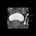
 in the right lobe and spontaneous subcapsular bleeding
  , a potential complication of hepatic adenomas.
 with central areas of rupture and hemorrhage
  . (Courtesy M. Yeh, MD, PhD.)
 with large peripheral vessels
  .
.
Stay updated, free articles. Join our Telegram channel

Full access? Get Clinical Tree


