KEY FACTS
Terminology
- •
Infection of humans caused by larval stage of Echinococcus granulosus or Echinococcus multilocularis
Imaging
- •
Best diagnostic clue: Membranes ± daughter cysts in complex heterogeneous mass
- •
E. granulosus
- ○
Anechoic cyst with double echogenic lines separated by hypoechoic layer
- ○
Honeycomb cyst, multiple septations
- ○
Water lily sign: Complete detachment of membrane
- ○
Snowstorm pattern: Anechoic cyst with internal debris and hydatid sand
- ○
- •
E. multilocularis
- ○
Single/multiple echogenic lesions
- ○
Irregular necrotic lesions with microcalcifications
- ○
Ill-defined infiltrative solid masses
- ○
Top Differential Diagnoses
- •
Hemorrhagic or infected cyst
- •
Complex pyogenic abscess
- •
“Cystic” metastases
- •
Biliary cystadenocarcinoma
Pathology
- •
Caused by larval stage of Echinococcus tapeworm
- ○
E. granulosus
- –
Most common form of hydatid disease, unilocular
- –
- ○
E. multilocularis
- –
Less common but aggressive
- –
- ○
Clinical Issues
- •
Serologic test positive in > 80% of cases
Diagnostic Checklist
- •
Rule out other complex or septate cystic liver masses
Scanning Tips
- •
Imaging clue: Daughter cysts can float freely within mother cyst
- •
Altering patient’s position may change position of daughter cysts
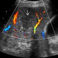
 with numerous peripheral daughter cysts, or scolices,
  within the right lobe of the liver.
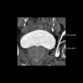
 . The fibrous rim
  can be seen surrounding the cyst. (From DP: Spleen.)
 and central heterogeneous content
  in the left lobe of the liver. Note the posterior acoustic enhancement
  .
.


