Fig. 6.1
Patient with HCC involving the right lobe of the liver as seen by the irregular hypodense lesion on the coronal section of CECT images (a) involving seg IV/VIII/VII and V and presence of right portal vein (PV) thrombosis, extending to MPV and SMV. FDG PET/CT done for staging shows intense FDG uptake in the primary mass (b) involving the lesion in right lobe of the liver with linear uptake (arrow) correlating with the tumor thrombus in the portal vein

Fig. 6.2
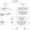
12 × 9 × 9 cm mass in the right lobe of the liver with thrombosis of the right and main portal vein as seen on the transaxial CECT images (a). The correlative transaxial images of the FDG PET/CT study show no significant FDG uptake in the liver lesion (b) suggestive of a tumor with good biology
HCC with metastases have a poor prognosis with limited treatment options, while locally advanced HCC in the absence of extrahepatic spread could be offered aggressive local therapies; thus, accurate staging helps triage patients. FDG PET has been useful in the detection of distant metastases of HCC and fares better than conventional imaging modalities for detection of bony involvement while showing similar detection rates for lung and nodal disease [6, 7] (Fig. 6.3).

Fig. 6.3
A case of intermediate HCC involving the right lobe of the liver treated with TACE and planned for TARE. FDG PET/CT images show FDG uptake in the residual disease within the large heterogeneous lesion in the right lobe of the liver (arrow in a, b). Also noted is the FDG uptake in the metastatic nodule in the right adrenal gland on the transaxial and coronal-fused PET/CT images (arrow head in c, d)
A systemic review and meta-analysis evaluating FDG PET or PET/CT in extrahepatic metastases and recurrent disease included eight studies and showed pooled estimates of sensitivity, specificity, positive likelihood ratio, and negative likelihood ratio of FDG PET (PET/CT) in the detection of extrahepatic metastases at 76.6% (95% CI, 68.7–83.3%), 98.0% (95% CI, 92.8–99.8%), 14.68 (95% CI, 5.5–39.14), and 0.28 (95% CI, 0.20–0.40), respectively [8].
Few studies which tried to evaluate its role as a biologic marker identified that tumors with a higher density of glucose receptors tend to be aggressive; hence, tumors which show greater FDG uptake could represent a disease with a bad biology [3, 9].
Tumors with no FDG uptake have better outcomes [10], while among FDG-avid tumors, those with higher FDG uptake show poor outcomes as compared with tumors with lower FDG concentrations. Tumors with a greater FDG concentration also tend to show a shorter doubling time and present with higher stage of disease [11–13].
6.1.2 Treatment Response Assessment
Local targeted treatment (LRT) for HCC exploits the pathophysiology of dual blood supply of hepatic tumors. It blocks the predominant arterial blood supply leading to reduction in blood flow and cell death via either ischemia, thermal/coagulation, or radiation effect which do not result in tumor shrinkage [14, 15], but show necrosis and reduction in the enhancement pattern which are not accounted in RECIST 1 or RECIST 1.1 criteria. The newer guidelines have incorporated the enhancement criteria (mRECIST) and necrotic parameters in the response assessment of HCC [16–18].
Identifying enhancement features could be difficult due to benign posttreatment inflammatory changes, or a heterogeneous nature of the tumor environment and functional imaging like diffusion-weighted magnetic resonance (DWMRI) or FDG PET/CT is recommended.
Response assessment with FDG PET/CT is done either by visual assessment of the tumor site in the pre- and post-therapy scans in comparison with blood pool uptakes or by calculating the reduction of FDG uptake at the tumor site using various semiquantitative or quantitative methods, e.g., ratio of tumor SUV to the liver or mediastinum or SUV max. Studies show better survival and event-free rates in patients who depict significant reduction in the uptakes at the tumor site [19, 20]. When compared to conventional imaging methods like CECT, FDG PET/CT showed a higher sensitivity in identifying residual viable tissue which is generally seen as a focal eccentric uptake in the periphery [21, 22] (Fig. 6.4).

Fig. 6.4
Case of HCC involving the right lobe of the liver with portal thrombosis, patient was planned for trans arterial radioembolization and hence referred for a staging FDG PET/CT study. Intense FDG uptake was seen in the tumor involving a large part of the right lobe with uptake also seen in the portal vein thrombosis as seen on the MIP (a) and the coronal fused images (arrow head in b). Increased uptake is seen in the thoracic region bilaterally on the MIP image (arrows in a) which corresponds to filling defect seen in the pulmonary vein in the coronal-fused images (arrow head in c). The CECT of the thoracic region in the coronal image confirms the presence of bilateral pulmonary thrombosis (arrow head in d)
In a bid to standardize the response assessment of solid tumors using FDG PET/CT, the PERCIST criteria was suggested by Wahl et al., which is adapted from the anatomical-based RECIST 1.1 principle and measures the FDG standard uptake (lean) in up to five index lesions (up to two lesions per organ) with highest FDG uptake. The response is expressed as percentage change in peak standard uptake of sum of lesions of baseline and posttreatment scans [23] (Table 6.1).
Table 6.1
Criterias for assesment of treatment response to conventional and targeted therapies (adapted from [24])
RECIST 1.1 | WHO | EASL | mRECIST | PERCIST | |
|---|---|---|---|---|---|
Complete response (CR) | Disappearance of all TL (up to 2 liver lesions) | Disappearance of all TL | Disappearance of all VL | Disappearance of all VL (up to 2 measurable liver lesions) | Disappearance of FDG uptake in the target lesions |
Partial response (PR) | ≥30% reduction in sum of greatest dimensional diameter of TL (compared to the baseline sum of diameter of TL) | ≥50% reduction in the sum of products of bidimensional diameter of TL (as compared to the baseline sum of diameters) | ≥50% reduction in the sum of products of bidimensional diameter of VL (as compared to the baseline sum of diameters) | ≥30% reduction in sum of greatest dimensional diameter of VL (compared to the baseline sum of diameters) | Minimum reduction of 30% of SUV (lean) in measurable target lesion |
Progressive disease (PD) | ≥20% increase in the sum of diameter of TL (compared to the smallest sum of diameter of TL recorded since treatment started) | ≥25% increase in the sum of diameter of TL (compared to the smallest sum of diameter of TL recorded since treatment started) | ≥25% increase in the sum of diameter of VL (compared to the smallest sum of diameter of VL recorded since treatment started) | ≥20% increase in the sum of diameter of VL (compared to the smallest sum of diameter of VL recorded since treatment started) | Increase of >30% of SUV (lean) or a new lesion identified on |
Stable disease | Any case that do not qualify for PR or PD | Any case that do not qualify for PR or PD | Any case that do not qualify for PR or PD | Any case that do not qualify for PR or PD | Any case that do not qualify for PMR or PMD |
The ideal time to assess response would be at 3 months post therapy allowing for posttreatment-related changes to settle which could cause false-positive or equivocal readings.
Radiofrequency ablation is a localized treatment option for smaller tumors and those away from vessels. FDG PET/CT for this indication should be performed prior to initiation of inflammatory changes, i.e., within 6–12 h of procedure to avoid masking of the residual disease seen as a focal uptake in the periphery [25] (Fig. 6.5).

Fig. 6.5
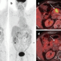
Hepatic metastasis in a case of colon carcinoma, FDG PET/CT study done for restaging revealed a solitary liver lesion as seen in the whole body maximum intensity projection (MIP) (a) and well appreciated on the fused transaxial image (b). Post RFA FDG PET/CT study shows a photopenic area on the coronal PET image (c) which corresponds to the site of lesion with no uptake within or in the periphery as seen in the fused image (d) depicting completeness of the procedure
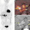
6.1.3 Utility of PET/CT in Evaluating Radioembolization of HCC
Post therapy scans in patients treated with 90 Yttrium tracers are evaluated with a bremsstrahlung imaging using the SPECT/CT scanner. Positron emissions from the Y90 radioisotope have been utilized to obtain an immediate post therapy PET/CT study. The advantage of this modality is the clear demarcation of region of radioisotope delivery and dosimetry to calculate actual dose delivered (Fig. 6.6). The presence of small extravasation of tracer into stomach or elsewhere is also identified which could have been missed on a pretreatment shunt evaluation dummy scan with colloids [26–28].

 Management of Hepatobiliary and Pancreatic Malignancies
 Hepatobiliary and Pancreatic Malignancies: Epidemiology, Clinical Presentation, Diagnosis, and Staging
 PET/CT in Pancreatic Malignancies
 Pathology of Hepatobiliary and Pancreatic Cancer
 Radiological Imaging in Hepatobiliary and Pancreatic Malignancies
 PET/CT in Gall Bladder and Biliary Tract Malignancies


Related posts:
Management of Hepatobiliary and Pancreatic Malignancies
Hepatobiliary and Pancreatic Malignancies: Epidemiology, Clinical Presentation, Diagnosis, and Staging
PET/CT in Pancreatic Malignancies
Pathology of Hepatobiliary and Pancreatic Cancer
Radiological Imaging in Hepatobiliary and Pancreatic Malignancies
PET/CT in Gall Bladder and Biliary Tract Malignancies
Stay updated, free articles. Join our Telegram channel
Full access? Get Clinical Tree
