KEY FACTS
Terminology
- •
Hepatocellular carcinoma (HCC)
- ○
Primary malignancy of liver arising from hepatocytes
- ○
- •
Synonyms: Hepatoma, primary liver cancer
Imaging
- •
Ultrasound: Primary test for screening/surveillance for HCC
- ○
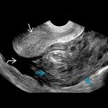
Solid, intrahepatic mass may be hypoechoic (most common), hyperechoic, or isoechoic
- ○
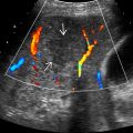
May see hypoechoic halo, mild posterior acoustic enhancement, or internal vascularity on color Doppler
- ○
Infiltrative tumors often difficult to visualize margins; look for refractive edge shadows, parenchymal distortion, or loss of normal echogenic portal triads, which are helpful secondary findings
- ○
Portal vein or hepatic vein tumor invasion highly suggestive of HCC
- ○
- •
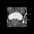
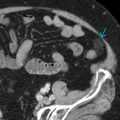
Contrast-enhanced CT, MR, or CEUS: For characterization
- ○
Arterial hyperenhancement and portal/delayed washout
- ○
Top Differential Diagnoses
- •
Intrahepatic cholangiocarcinoma
- •
Regenerative or dysplastic nodule
- •
Hepatic hemangioma (especially hyalinized subtype)
- •
Focal nodular hyperplasia
- •
Hepatic adenoma
- •
Metastases
Clinical Issues
- •
Screening/surveillance ultrasound every 6 months recommended for the following populations in USA
- ○
Cirrhosis (60-90%) from any cause
- –
Most commonly due to chronic viral hepatitis (HBV, HCV), alcoholic cirrhosis
- –
- ○
Noncirrhotic chronic Hepatitis B in certain populations
- –
Asian male > 40, Asian female > 50, African/North American black, family history HCC
- –
- ○
Surveillance benefit uncertain in following populations: Noncirrhotic chronic HCV with stage 3 fibrosis, noncirrhotic nonalcoholic fatty liver disease, and noncirrhotic HBV carriers with active hepatitis who do not meet above criteria
- ○
Scanning Tips
- •
Careful inspection of portal veins and hepatic veins for thrombus critical; infiltrative HCC often invades veins
- •
Liver dome lesions are often missed; angle transducer to image above HV to completely visualize liver dome
- •
RT and LT inferior tip of liver lesions are easily missed; carefully scan through very inferior portions of liver
- •
Anterior lesions often poorly visualized with curvilinear or vector transducers; diligent use of 9-MHz linear probe helps visualization of superficial portions of liver
- •
Trying multiple probes, positions, and windows are key to improve detection; some liver lesions may only be seen on left lateral decubitus position (vs. supine) or intercostal approach (vs. subcostal)
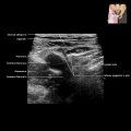
 . Numerous adjacent satellite nodules
  and portal vein invasion
  are depicted. Underlying liver disease is evident given the nodular liver capsule
  and ascites
  .
.Related posts:

Stay updated, free articles. Join our Telegram channel

Full access? Get Clinical Tree


