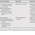
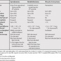
55 The differential diagnosis in the setting of chronic diffuse interstitial lung disease relies upon recognition of specific high-resolution computed tomography (HRCT) signs. These include interlobular septal thickening, reticular densities, cystic air spaces, nodules, ground-glass attenuation, and consolidation. In normal individuals, a few thin interlobular septa may be visualized on HRCT in the subpleural regions. When multiple contiguous septa become thickened, a characteristic appearance of polygonal arcades can be seen. Septal thickening may be due to fluid, cellular infiltration, fibrosis, or a combination of these. Smooth septal thickening favors pulmonary edema over lymphangitic carcinomatosis. Nodularity or beading of the septa favors the diagnosis of lymphangitic carcinomatosis. Irregular septal thickening implies the presence of underlying fibrosis and may be seen in sarcoidosis, asbestosis, and idiopathic pulmonary fibrosis.
High-Resolution Computed Tomography of Chronic Diffuse Infiltrative Lung Disease
Septal Thickening
Reticular Densities
Stay updated, free articles. Join our Telegram channel

Full access? Get Clinical Tree


