Hodgkin Lymphoma


Hodgkin Lymphoma


Stephanie A. Terezakis

Melissa M. Hudson

Louis S. Constine


Pediatric Hodgkin lymphoma (PHL) presents a challenge to continue making progress in its curability while diminishing the risk for the morbidities that compromise quality of life and survival. The history of PHL is, of course, the history of Hodgkin lymphoma (HL). In 1832, Thomas Hodgkin (1) described seven patients with enlarged absorbent (lymphatic) glands not thought to result from inflammation. At the turn of the century, Sternberg and Reed each described the multinucleated giant cell characteristic of HL (2). Shortly after the discovery of x-rays in 1902, Pusey (3) demonstrated the radioresponsiveness of HL. In the 1930s, Gilbert (4) laid the foundation for its definitive treatment with radiotherapy (RT), and Peters (5) provided additional definition of important principles. During World Wars I and II, the lympholytic effects of nitrogen mustard were recognized (6), and over the next two decades progress in safely combining multiple chemotherapeutic agents to treat HL led to DeVita’s (7) report on the use of nitrogen mustard, vincristine (Oncovin), procarbazine, and prednisone (MOPP) chemotherapy. Kaplan (2) systematically studied the role of RT for HL during these decades. Concurrently, advances were made in identifying different pathologic subtypes, determining staging criteria, improving diagnostic imaging capabilities, and developing effective chemotherapeutic regimens. When the clonality of the Hodgkin-Reed-Sternberg (HRS) cell was finally established in the 1960s, controversy over the malignant or inflammatory nature of HL abated (8).

Although the biology and natural history of HL in children are similar to that in adults, when irradiation techniques and dosages suitable for controlling disease in adults were translated to the pediatric setting, substantial morbidities (primarily musculoskeletal growth inhibition) were produced (9,10). It is in this context the new strategies for the treatment of pediatric HL were developed by Donaldson and others (11, 12, 13, 14, 15, 16, 17, 18, 19). Historically, children were thought to have a worse prognosis than adults (2). It is now apparent that the converse is true (20, 21, 22).


BIOLOGY

For many years, the rarity and distribution of malignant cells in pathologic material precluded elucidation of the origin and evolution of the HRS cell. Advances in immunohistology and molecular biology subsequently revealed that the HRS cell and its variants most commonly derive from a neoplastic clone that originates from B lymphocytes in lymphoid germinal centers. Clonality can be demonstrated at diagnosis and relapse through detection of unique nucleotide sequences, which molecularly fingerprint the HRS cell clone; these sequences represent rearrangements of immunoglobulin variable-region (V) genes, which are B-cell derived (23,24). Complicating this scenario is the finding by Kanzler et al. (24) that flawed V genes, which would be lethal for normal B cells, are present in HRS cells and prevent them from expressing immunoglobulin. Such cells would be expected to die of apoptosis. Therefore, the genesis of classic HL (CHL) probably involves evasion by the HRS cell of the apoptotic pathway. Deregulation of the nuclear transcription factor (NF-κB) in the HRS cells has been hypothesized as a mechanism that prevents apoptosis (25). Epstein-Barr virus (EBV) and genes that monitor cell damage, such as p53, may play a role in the rescue and repair of the HRS cells (24,26). EBV genome fragments can be found in HRS cells in 30-50% of HL specimens, most commonly in the mixed-cellularity HL (MCHL) subtype and rarely in the lymphocyte-predominant HL (LPHL) (27,28). The EBV genome is temporally stable because it can be found at diagnosis and relapse. Finally, the HRS cell can also have characteristics of T lymphocytes and the interdigitating reticulum cell. Such evidence of a multilineage origin of the HRS cell may be explicable by postulating that the HRS cell is a hybridoma resulting from fusion of different cell lines, provoked by a virus or other agent. Cytogenetic data also show an unexpected frequency of B-cell translocation (14:18) and bcl-2 gene involvement in HL, but these may derive from bystander normal lymphocytes rather than the HRS cell (29). The complexity of elucidating the etiology of HL continues to unfold, and is illustrated by recent data that not only support the association of infectious mononucleosis with EBV-positive HL, but also suggest that EBV-negative HL has a different etiology (30). A pathogenetic model for HL is depicted in Figure 7.1, which suggests that the HRS cells arise in a germinal lymphoid center from a clone of antigen-stimulated B cells and through genetic changes achieve immortality and malignant properties (31).

A curious characteristic of HL is the rarity (about 1%) of the malignant HRS cell in specimens and the abundant reactive cellular infiltrate of lymphocytes, macrophages, granulocytes, and eosinophils. The histologic features and clinical symptoms of HL have been attributed to the numerous cytokines secreted by the HRS cells, which include interleukin (IL)-1 and IL-6, and tumor necrosis factor (TNF) (32,33). IL-5 could be responsible for the eosinophilia in MCHL, and transforming growth factor β could be responsible for the fibrosis in the nodular sclerosis subtype. These cytokines also enable the cells to evade immunologic surveillance and promote their own replication (32).

Evidence that HL comprises a family of diseases includes the observation that HRS cells can rarely be
derivatives of cytotoxic T cells and that nodular lymphocyte-predominant HL (NLPHL) is a distinctive and uncommon lymphoproliferative disorder. The lymphocytic and histiocytic (L&H) cells characteristic of NLPHL have folded and lobate nuclei among small lymphocytes and histiocytes. The L&H cells are also B cell-derived but harbor V gene alterations, which differ from those in HRS cells; whether the L&H cells are monoclonal is unclear (34,35). The clinical characteristics of NLPHL differ from those of the subtypes of CHL by virtue of its indolence, excellent prognosis, epidemiology, and response to chemotherapy. The Revised European-American Classification of Lymphoid Neoplasia (REAL) has devised a schema that reflects the distinction of NLPHL from CHL (36).


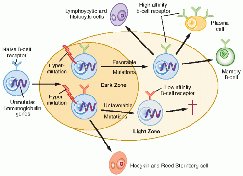
Figure 7.1 The derivation of Hodgkin and Reed-Sternberg cells in classic Hodgkin’s lymphoma and lymphocytic and histiocytic cells in nodular lymphocyte-predominant Hodgkin’s lymphoma. (From Thomas RK, Re D, Wolf J, et al. Part 1: Hodgkin’s lymphoma- molecular biology of Hodgkin and Reed-Sternberg cells. Lancet Oncol 2004;5:11-18.)

Several aspects of the biology of HL continue to unfold, and many have prognostic applicability. For example, loss of CD 15 expression may be a prognostic factor for PHL clinical outcomes, although CD 15 expression has been inconsistent possibly due to biologic or technical variability (37,38). A high proliferative index (PI) may suggest chemoresponsiveness and, as a result, a low pre-treatment PI may be associated with poor outcomes (37,38).


EPIDEMIOLOGY

HL makes up 6% of childhood cancers. A striking male:female predominance is found among children, with a ratio of 4:1 for 3- to 7-year-olds, 3:1 for 7- to 9-year-olds, and 1.3:1 (a ratio more similar to that of adults) for older children (20,39,40). The age-specific incidence curves for HL in the United States and other developed countries are bimodal, peaking in the 20s and then again after the age of 50 (41). The disease is uncommon

before age 5 and, among children, is most common in adolescence. Not only does the incidence of PHL vary in younger children versus adolescence, but the histologic subtypes and associations with EBV also vary (42).

Although evidence that HL is infectious or contagious was suggested by reports of case clusters (43), confirmatory data are lacking. However, the role of EBV in its pathogenesis of EBV-positive HL is well established (30). EBV early RNA1 is expressed in HRS cells in 58% of childhood cases, most commonly in those with mixed-cellularity histology (44). Of particular interest, expression was age-dependent: 75% of children under the age of 10 years compared with 20% of older children. In addition, a history of infectious mononucleosis increases the risk for HL, and anti-EBV titers are elevated before diagnosis of HL.

In a large population-based study, infectious mononucleosis was associated with an increased risk of EBV-positive HL with an odds ratio of 3, but did not increase risk for EBV-negative HL (30). EBV latent membrane protein (LMP-1) is expressed in CHL and has been correlated with serum IL-10 levels (45). LAG-3 expression, a marker for regulatory T cells, is also associated with EBV positivity and related to impairment of LMP-1 function (46). Several reports have suggested that EBV positivity is most common in the MCHL subtype (47, 48, 49). There has been conflicting evidence as to whether latent EBV infection carries prognostic significance. LMP-1 positivity has been associated with inferior survival in patients with nodular sclerosis cellular subtype Bennett II and those with advanced-stage disease despite a lack of influence on failure-free survival. LMP-1 emerged as an independent prognostic factor for overall survival (OS) on multivariate analysis in a study by Claviez et al. Latent EBV infection has also been
associated with inferior OS in patients greater than the age of 50 years when adjusting for stage, sex, and B symptoms, although it was not associated with inferior disease-specific survival (48). Analysis of the database of the International Hodgkin Study Group revealed no significant differences associated with EBV positivity in failure-free survival or OS after adjustment for stage and age (45,49). Understanding the pathogenesis and prognostic implications of EBV-associated HL may help to improve immune-directed therapeutic strategies.

Evidence of a genetic predisposition exists and is relevant when counseling families. The incidence of HL is two to five times higher in the siblings of affected children and nine times higher in same-sex siblings than in the general population. Parent-child associations have been reported (41,50). Mack et al. (51) reported a 99-fold increased risk in monozygotic twins of patients but no increased risk in dizygotic twins. The status of the immune system in patients with HL also deserves comment. A complex deficiency in cellular immunity exists, which includes a deficiency in naive T cells and an elevated sensitivity of effector T cells to suppressor monocytes and T-suppressor cells (52). Of interest is that radiation results in a long-term dysregulation of T-cell subset homeostasis. Finally, it is unclear whether HL is more common in patients with either congenital (e.g., ataxia-telangiectasia) or acquired immunodeficiency states, including acquired immunodeficiency syndrome (43,53), but HL is rarely seen as a second malignancy. In patients with the human immunodeficiency virus, the disease more commonly presents in an advanced stage with systemic symptoms, extranodal involvement, and a poor response to therapy (54).


Table 7.1 Ann Arbor Staging System with Cotswold Modifications for Hodgkin Lymphoma


Stage


Description


I


Involvement of a single lymph node region or lymphoid structure (e.g., spleen, thymus, the Waldeyer ring, or single extralymphatic site [IE])


II


Involvement of two or more lymph node regions on the same side of the diaphragm or localized contiguous involvement of only one extranodal organ or site and lymph node region on the same side of the diaphragm (IIE). The number of anatomic sites is indicated by a subscript (e.g., II3)


III


Involvement of lymph node regions on both sides of the diaphragm (III), which may be accompanied by involvement of the spleen (IIIs) or by localized contiguous involvement of only one extranodal organ site (IIIE) or both (IIISE)


III1


With or without involvement of splenic hilar, celiac, or mesenteric nodes


III2


With involvement of para-aortic, iliac, or mesenteric nodes


IV


Diffuse or disseminated involvement of one or more extranodal organs or tissues, with or without associated lymph node involvement


Designations applicable to any stage


A


No symptoms


B


Fever (temperature >38°C), drenching night sweats, unexplained loss of >10% of body weight in the preceding 6 months


X


Bulky disease (a widening of the mediastinum by more than one third or the presence of a nodal mass with a maximal dimension >10 cm)


E


Involvement of a single extranodal site that is contiguous or proximal to the known nodal site


CS


Clinical stage


PS


Pathologic stage (as determined by laparotomy)


Modified from Kaplan H. Hodgkin’s Disease. Cambridge, MA: Harvard University Press; 1980; and Grufferman S, Delzell E.


Epidemiology of Hodgkin’s disease. Epidemiol Rev. 1984;6:76-106, with permission.


CLINICAL PRESENTATION

HL appears to be unifocal in origin, with 90% of patients presenting in a pattern that suggests contiguous lymphatic spread (2,55). Most children are diagnosed on the basis of supradiaphragmatic lymph nodes, with painless cervical adenopathy in 80%. The nodes are generally firm and may be tender. Mediastinal involvement occurs in 76% of adolescents but in only 33% of 1- to 10-year-olds. Mediastinal disease may produce symptoms such as dyspnea, cough, and superior vena cava syndrome. Axillary adenopathy is less common (2). Associations exist between the mediastinum and the neck, the neck and the ipsilateral axilla, the mediastinum and the hilum, and the spleen and abdominal lymph nodes (55). Isolated mediastinal or infradiaphragmatic HL is rare, occurring in fewer than 5% of patients. About one third of the patients have systemic “B” symptoms, as defined in Table 7.1 (2,56).


PATHOLOGIC CLASSIFICATION

The HRS cell is the essential malignant cell in HL (Fig 7.2). It is large, with abundant cytoplasm, two or three nuclei, and a prominent nucleolus. However, its frequency in pathologic specimens is greatly variable because of the presence of numerous reactive cells including lymphocytes, eosinophils, and plasma cells. Moreover, the HRS cell, particularly its mononuclear variant, is not pathognomonic for HL because cells simulating it can be found in other disorders that are reactive, infectious, or malignant (57). The diagnosis of HL must be established by lymph node biopsy. Aspiration cytology alone is not recommended because of the lack of stromal tissue, the
small number of cells present in the specimen, and the difficulty of classifying HL into one of the four categories of the Rye classification. The Rye classification subcategorizes HL into nodular sclerosing (NSHL), MCHL, LPHL, and lymphocyte-depleted (LDHL) types. With modern treatment the prognostic significance of these subtypes has diminished, although the presenting characteristics and natural history remain evident, particularly for the nodular subtype of LPHL. The importance of distinguishing NLPHL from the other types has led to the REAL classification (Table 7.2) (39). The clinicopathologic characteristics are the same as those in adults and are briefly described here.


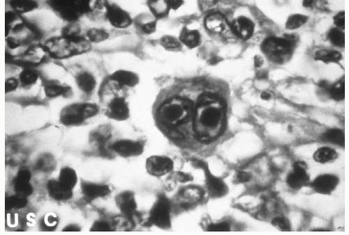
Figure 7.2 Reed-Sternberg cell.


  • NLPHL (58): The distinctive cell is the L&H “popcorn” cell, which is CD20 (B-lymphocyte marker) positive and CD 15 negative. Classic HRS cells (which are usually CD15 and CD30) are rare, as is the detection of EBV. Progressive transformation of the germinal centers of lymph nodes is often seen, and, in fact, can occur in the absence of NLPHL. Therefore, it is important to distinguish these entities. NLPHL has a long natural history, in its time to diagnosis and to relapse, reminiscent of that of indolent non-Hodgkin lymphomas (NHL). It is more common in young children (33% of all patients are younger than 15 years), has a high male:female ratio (4:1), and commonly involves a single lymph node region with sparing of the mediastinum (59).


  • Lymphocyte-rich (classic) HL: HRS cells (CD15) are identifiable against a background predominantly of lymphocytes. Clinical behavior is similar to that of MCHL.


  • Mixed-cellularity (classic) HL: HRS cells (CD15) are common against a background of abundant normal reactive cells (lymphocytes, plasma cells, eosinophils, histiocytes). This subtype can be confused with peripheral T-cell NHL. MCHL is less common in children, often accompanied by “B” symptoms, and more often involves infradiaphragmatic nodes.


  • Nodular sclerosis (classic) HL: This subtype is distinctive because of the presence of collagenous bands that divide the lymph node into nodules, which often contain an HRS cell variant called the lacunar cell. NSHL often occurs in children, involves supradiaphragmatic nodes, and spreads in an orderly manner along contiguous nodal chains.


  • Lymphocyte-depleted (classic) HL: This subtype is rare and commonly confused with NHL, particularly of the anaplastic large cell type. HRS and pleomorphic variants are common relative to the number of background lymphocytes. LDHL often is advanced at diagnosis and has a poor prognosis.


Table 7.2 Comparison of Revised European-American Classification of Lymphoid Neoplasia (REAL) and Rye Classification of the Hodgkin Disease


REAL Classification


Rye Classification


Lymphocyte predominance, nodular


Lymphocyte predominance, nodular (most cases)


Classic Hodgkin disease


Lymphocyte-rich


Lymphocyte predominance, diffuse (most cases)


Nodular sclerosing


Lymphocyte predominance, nodular (some cases)


Mixed cellularity


Mixed cellularity


Lymphocyte depletion


Lymphocyte depletion


Modified from Klein G. Epstein-Barr virus-carrying cells in Hodgkin’s disease. Blood. 1992;80(2):299-301, with permission.


Although the pathologic and immunohistochemical characteristics of HL generally are sufficiently clear to establish a diagnosis, confusion with select subtypes of NHL is problematic. In particular, CHL and NLPHL can be confused with anaplastic large cell NHL, and LPHL can be confused with T cell-rich B-cell NHL (32,39). NHL, and LPHL can be confused with T cell-rich B-cell NHL (32,39).

The relative distribution of the subtypes in younger children differs from that in adolescents and adults, as reported from Stanford University (20). LPHL is more common (13%) in younger children (younger than 10 years), whereas LDHL is exceedingly rare. Although NSHL is the most common subtype in all age groups, it is more common in adolescents (77%) and adults (72%) than in younger children (44%). Conversely, MCHL is more common in younger children (33%) than in adolescents (11%) or adults (17%).


STAGING

Using anatomic groups of regional lymph nodes, the staging system was designed for all age groups, according to a modification of the system devised at the 1970 Ann Arbor Symposium. This was subsequently revised at the Cotswold’s Meeting, although not all suggestions from those recommendations are consistently used (56). In this system patients are assigned a clinical stage, and if staging laparotomy is performed, the patient is assigned a pathologic stage (Table 7.1) (2).

A weakness of the Ann Arbor system is its failure to consider disease bulk (either dimension or number of involved sites) or specific patterns of involvement. For this reason,
subclassifications of the Ann Arbor staging system have been proposed, particularly for patients with large mediastinal adenopathy (LMA) or stage IIIA disease. LMA, most commonly defined as a mass exceeding one third the transverse diameter of the chest (intrathoracic width measured at the dome of the diaphragm) on a standard upright posteroanterior chest radiograph, places a patient at a greater risk for disease recurrence after radiation alone. Patients with pathologic stage (PS) III disease limited to the spleen or splenic, celiac, or portal nodes are denoted anatomic substage III1 and considered to have a more favorable prognosis than patients with involvement of paraaortic, iliac, or mesenteric nodes, denoted as III2 (60). However, this system has not proven useful in some centers.

The distribution of stages observed in children is somewhat different from that observed in adults. Table 7.3 summarizes the demographic and clinical features of children, adolescents, and young adults presenting with HL (61). Among 3571 consecutive patients with HL treated at three pediatric centers, 18.1% were younger than 10 years and 81.4% were 11-16 years old. Stage I or II disease was present in 65.8% of children.


Table 7.3 Demographic and Clinical Characteristics at Presentation of Pediatric Hodgkin Disease


Age Range


Childhood HL ≤14 years


AYA HL 15-35 years


Adult HL ≥35 years


Older Adult HL ≥ 55 years


Prevalence of HL


10-12%


50.00%


35.00%


Cases


Gender


Male:Female


2-3:1


1:1-1.3:1


1.2:1-1:1.1


Histology


Nodular sclerosis


40-45%


65-80%


35-40%


Mixed cellularity


30-45%


10-25%


35-50%


Lymphocyte-depleted


0-3%


1-5%


2-6%


NLPHL


8-20%


2-8%


7-10%


EBV-associated


27-54%


20-25%


34.00-40%


50-56%


Risk factors: male, younger age, mixedcellularity histology, economically disadvantaged countries


Other risk factors


Lower SES


Higher SES


Increasing family size


Smaller family size


Early birth order


Stage at presentation


30-35 % with stage III or IV disease


40% with stage III or IV disease


55% with stage III or IV disease


25% with B symptoms


30-40% with B symptoms


50% with B symptoms


Trend to less bulky disease, less mediastinal mass but more advanced stage with higher IPS scores


Relative survival rates at 5 years


94% (<20 years)


90% (<50 years)


65% (>50 years)


SES, socioeconomic status


From Punnett A, Tsang R, Hodgson DC. Hodgkin lymphoma across the age spectrum: epidemiology, therapy, and late effects.


In: Constine, LS, ed. Cancer genesis, treatment, and late effects across the age spectrum. Seminars in Radiation Oncology. 2010;20(1):30-44.


DIAGNOSTIC EVALUATION

After pathologic confirmation, the patient undergoes an extensive clinical staging. This begins with a detailed history of systemic symptoms and evidence of cardiorespiratory compromise or organ dysfunction. The physical examination carefully records the location and size of all palpable lymph nodes. An evaluation of Waldeyer ring, cardiorespiratory status, and organomegaly is vital. Laboratory studies include complete blood count with platelets and biochemical evaluation of renal and liver function. Acute phase reactants, including erythrocyte sedimentation rate, serum copper, and ferritin, may be elevated at diagnosis and can be used as nonspecific marker of disease activity. Elevated serum CD30 and CD25 have been correlated with advanced stage, the presence of constitutional symptoms, and poor prognosis; however, these studies have not been widely used to stage and monitor patients during therapy (62,63). C-reactive protein, another acute phase protein produced in the liver, holds promise as a diagnostic and prognostic index for both Hodgkin and cardiovascular
diseases (64). Patients with “B” symptoms or stage III-IV disease should undergo bone marrow biopsy (65). The low yield of a bone marrow evaluation in asymptomatic patients with localized disease (stage I and II) does not support routine use of the procedure.

Imaging studies of the thorax should include a chest radiograph with a posteroanterior and lateral view. It should be noted that the LMA ratio, if based on computed tomography (CT) scan measurements, would increase the frequency of patient’s LMA since a mediastinal mass will splay bilaterally in a reclined child. CT scans are essential for delineation of the status of intrathoracic lymph node groups (including the hila and cardiophrenic angle), lung parenchyma, pericardium, pleura, and the chest wall, demonstrating abnormalities in about one half of patients with unremarkable chest radiographs (22,66). Definition of disease involvement of intrathoracic tissues by CT often dictates more aggressive therapy than would otherwise have been administered. Distinguishing normal (or hyperplastic) thymus from nodes in children can be problematic.

Imaging of the abdomen and pelvis may involve an abdominal and pelvic CT scan or magnetic resonance imaging (MRI). If CT is used, oral and intravenous contrast administration is needed to accurately distinguish retroperitoneal and pelvic lymph nodes from other infradiaphragmatic structures. In cases with suboptimal contrast resolution of the bowel, MRI may provide a better evaluation of the fat-encased retroperitoneal nodes (67). In previous German and Pediatric Oncology Group (POG) trials using CT or MRI for staging, the size of abdominal and pelvic lymph nodes was used to define the disease involvement. Abdominal nodes less than 1.5 cm in diameter and pelvic nodes less than 2 cm were considered negative, whereas abdominal nodes more than 2 cm in diameter and pelvic nodes more than 3 cm were considered positive. Nodal biopsies were undertaken in equivocal nodes falling in between these sizes.


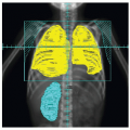
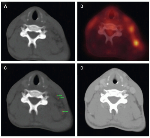
Figure 7.3 A: A few poorly identifiable lymph nodes in the left cervical area on an axial CT slice without IV contrast before chemotherapy. B: FDG-PET image of the same area. C: Measurements of the largest lymph nodes (3.7, 4.6 and 11.1 mm). D: The left cervical area after chemotherapy.

HL involving the liver and spleen is suggested by CT or MRI findings of organomegaly with areas of abnormal density. Organ size alone does not reliably predict lymphomatous involvement because tumor deposits may be less than 1 cm in diameter and not visualized by diagnostic imaging studies. In a POG analysis of 216 children, intrinsic spleen lesions and abnormal portohepatic and celiac nodal areas were highly predictive CT findings but were infrequently observed (68). Therefore, only histologic assessment can definitively evaluate the liver and spleen, but the indications for surgical staging of these organs are limited.

Nuclear imaging with gallium-67 was widely used to stage and monitor treatment response in children with HL. However, gallium has limitations because of its low resolution and physiologic biodistribution, which cause difficulties in evaluating abdominal and pelvic lymph nodes and necessitates delayed imaging. Positron emission tomography (PET) is now recognized as an integral staging modality for lymphoma (Fig. 7.3) (69, 70, 71). Uptake of the radioactive glucose analog fluorodeoxyglucose (FDG) correlates with proliferative activity in tumors undergoing anaerobic glycolysis. FDG-PET has advantages over gallium-67 because the scan is a 1-day procedure with higher resolution, better dosimetry, and less intestinal activity, has quantitation potential, and can provide better anatomic localization when combined with CT scan. Gallium-67 and FDG-PET both provide full-body imaging in which areas of abnormal avidity have been correlated with treatment outcomes. Both also have limitations in the pediatric setting (e.g., tracer avidity in nonmalignant thymic
rebound commonly observed after completion of therapy for HL).

Improved sensitivity and specificity of FDG-PET relative to CT/MRI and gallium scanning in PHL have been noted. Although FDG-PET and gallium were similar for staging, both response assessment and relapse identification were improved with FDG-PET in a study by Mody et al. (72). PET and gallium scans were performed at diagnosis, at early response assessment, and off therapy in HL patients at St. Jude Children’s Research Hospital. PET had increased sensitivity relative to gallium. PET upstaged four patients and changed response assessment in two patients although these findings only changed management in one patient. Gallium scanning did not change stage in any patient. PET and gallium scanning sensitivity were similar in the neck and mediastinal regions at diagnosis but the sensitivity of gallium is limited in infradiaphragmatic regions due to decreased resolution as a result of gallium excretion in the bowel. Neither positive PET nor gallium scan at early response predicted relapse. In this study, the negative predictive value (NPV) for PET at the end of therapy was 89.3% compared to 83% for gallium while the positive predictive value for PET was 28.6%. The predictive power of PET scans for early response and end of therapy evaluation requires further investigation with larger pediatric cohorts.


PROGNOSTIC FACTORS

As the treatment of HL has improved, characteristics that influence outcome have diminished in importance. However, several factors continue to influence the success and certainly the choice of therapy. These factors are interrelated in the sense that disease stage, bulk, and biologic aggressiveness often are codependent. Also complicating the determination of prognostic factors is that relevant variables often depend on staging evaluation and treatment. Most data are based on reports that include primarily adults.

The disease stage persists as the most important prognostic variable. Patients with advanced-stage disease, especially stage IV, have a worse outlook than those with early-stage disease (73).

The bulk of disease is reflected by the disease stage and is determined more specifically by the volume of distinct areas of involvement and the number of disease sites. LMA places a patient at a greater risk for disease recurrence. While OS remains high due to the effectiveness of salvage chemotherapy (2,22,74), patients with LMA have a somewhat lower survival rate in most, but not all series (2,22,75). Patients (at least those staged only clinically) with several sites of involvement, generally defined as more than three sites, fare less well (22). Patients with stage IV disease who have multiple organs involved fare especially poorly.

Nonwhite pediatric patients presented more frequently with advanced-stage disease based on a study population derived from two South African hospitals although white children had a lower OS (76). In this report, nonwhite pediatric patients had better survival when compared to white pediatric patients of the same stage. However, in a recent study from St. Jude Children’s Research Hospital, black pediatric patients had lower event-free survival (EFS), but equivalent 5-year OS to white pediatric patients (77). The impact of race on prognosis remains unclear particularly given the potential impact of socioeconomic, cultural, and environmental differences between white and black populations that may play a role in clinical outcomes.

Systemic (“B”) symptoms, which result from cytokine secretion, reflect biologic aggressiveness and confer a worse prognosis. The constellation of symptoms appears to be relevant to this observation. That is, patients with night sweats only (at least among patients with PS I and II disease) appear to fare as well as patients with PS I-IIA, and those with both fever and weight loss have the worst prognosis (78).

Laboratory studies/biologic markers, including the erythrocyte sedimentation rate, serum ferritin, hemoglobin level, serum albumin, and serum CD8 antigen levels, have been reported to predict a worse outcome (62,63,78,79). This could reflect disease biology or bulk. Other investigational serum markers associated with an adverse outcome include soluble vascular cell adhesion molecule-1 (80), TNF (81), soluble CD (28,82), β2-microglobulin (83), transferrin, and serum IL-10 (84). High levels of caspase 3 in HRS cells have been correlated with a favorable outcome (85). Serum vascular endothelial growth factor (VEGF) levels may also have prognostic implications. VEGF is a pro-angiogenic molecule involved in neovascularization of tumors. Increased microvessel density associated with increased VEGF level expression has been demonstrated in HL progression (86) and VEGF expression has been identified in HRS cells (87). Pretreatment levels of angiogenic factors have been found to be significantly elevated in HL patients compared to controls and high levels of VEGF have been associated with poor survival (88). Serum VEGF levels have also been associated with treatment response in children with HL (89). In a study by Ben Arush et al., serum VEGF levels correlated with treatment response in seven of nine children with HL. A significant reduction in serum VEGF level was identified in patients with a partial or complete response based on an assessment using PET-CT. The identification of angiogenic factors that play a role in the clinical course of HL may have therapeutic implications for the use of anti-angiogenic agents in the future. Other biologic factors that may enable prognostic modeling continue to be discerned (90,91). A recent study from the Groupe d’Etude des Lymphomes de l’Adulte (GELA) prospectively analyzed the prognostic value of TNF, soluble receptors TNF-R1 and TNF-R2, IL-10, IL1-RA, IL-6, and sCD30 in patients with an initial diagnosis of CHL. Five-year EFS probability was predicted by segregating combinations of serum marker levels into four prognostic classes, suggesting that a plasma cytokine signature may predict clinical outcomes in CHL (91).

Histologic subtype is relevant but may differ in children versus adults. For example, adults with clinical stage I or II MCHL have a higher frequency of subdiaphragmatic relapse, and which (indirectly) influences survival (22). Grade 2 NSHL histology has conferred poor outcome in some but not all studies (92). Patients with LDHL fare poorly. A recent report from the United Kingdom Children’s Cancer Study Group assessing the relevance of histology in 331 children is revealing. Fewer than 1% had LDHL, obviating any meaningful assessment of its prognostic significance. For patients with other histologies treated with combined therapy, no difference in outcome was observed (93). As previously discussed, patients with NLPHL have distinctive differences in disease-free survival and OS.

Age is a significant prognostic factor in some studies. Survival rates for children with HL approach 95%. In a report from Stanford, the 5- and 10-year survival rates for children younger than 10 years with HL are 94% and 92%, respectively, compared with 93% and 86% for adolescents (11-16 years old)
and 84% and 73% for adults (20). Although children younger than 4 years with HL are uncommon, even these children appear to have an excellent prognosis (21).

The rapidity of response to initial therapy is an important prognostic variable in many forms of cancer, including HL. In some trials, the rapidity of response to chemotherapy is used to determine subsequent therapy (94). Early response to therapy as measured by gallium-67 or FDG-PET imaging is under investigation as a possible marker of prognosis. The NPV of PET was recently evaluated in the GHSG HD15 trial. After patients received BEACOPP-based chemotherapy, PET was used to assess residual disease measuring greater than or equal to 2.5 cm in diameter. Progression-free survival was significantly improved in PET negative patients (96%) as compared to PET positive patients (86%) (p = 0.011). Although survival outcomes are not yet available due to the length of follow-up, response-adapted therapy proved promising with an NPV of 94% for PET in this study (95). In the setting of stem cell transplant for refractory/recurrent HL, pretransplant functional imaging response has been found to predict outcome. Patients with positive PET or gallium scans prior to undergoing high-dose chemotherapy with autologous stem cell transplant had a significantly lower 3-year PFS and OS rates than patients with negative functional imaging (96).

Although prognostic factors will continue to be influenced by choice of therapy, parameters such as disease, bulk, number of involved sites, and systemic symptoms are likely to remain relevant to outcome. Nonetheless, as therapy becomes increasingly tailored to prognostic factors and therapeutic response, overall outcome should become less affected by those parameters.


SELECTION OF THERAPY

HL is one of the pediatric malignancies that has an adult counterpart with a similar natural history and biology. However, devising the optimal therapeutic approach for children with this disease is complicated by their greater risk for adverse effects. In particular, RT dosages and fields used in adults can cause profound musculoskeletal retardation, including intraclavicular narrowing, shortened sitting height, decreased mandibular growth, and decreased muscle development in the treated volume (see Chapter 19) (9,10). Therefore, whereas adults with early-stage HL were historically treated with full-dose radiation as a single modality (22), in prepubertal children, despite a similar success rate, this approach produced unacceptable sequelae (10,19,74,97). Additionally complicating the treatment of children are sex-specific differences in chemotherapy-induced gonadal injury. The desire to cure young children with minimal side effects has stimulated attempts to reduce staging procedures, the intensity and types of chemotherapy, and the radiation dosage and volume. Because of the differences in the age-related developmental status of children and the sex-related sensitivity to chemotherapy, no single treatment method is ideal for all children.

Important studies using chemotherapy and low-dose radiation in children to effect cure with tolerable sequelae were first undertaken at Stanford University. These trials used chemotherapy in combination with lower dosages of RT for young children with early-stage disease (2,11,12). Although growth deformity was decreased, full-course MOPP is associated with a high risk of sterility and secondary leukemia (discussed in Chapters 19 and 20). Therefore, alternative regimens were used subsequently (14,16,51,98).

An important sequence of studies performed by the German-Austrian Pediatric Hodgkin’s Disease Study Group progressively refined the extent of staging and the intensity of chemotherapy and radiation (14,16,98). The nature and results of these studies (HD-82, HD-85, HD-87, HD-90, and HD-95) are summarized in Tables 7.4 and 7.5. Stage-dependent chemotherapy and radiation dosage and volume were used and these studies served as the paradigm for the risk-adapted treatment approach that is currently used at most pediatric treatment centers.

In general, the use of radiation and chemotherapy broadens the spectrum of potential toxicities while reducing the severity of individual (drug- or radiation-related) toxicities. The volume of radiation and the intensity and duration of chemotherapy are risk adapted, or determined by prognostic factors at presentation, including presence of constitutional symptoms, disease stage, and bulk. Results for patients with early (favorable) and advanced (unfavorable) HL are summarized in Tables 7.4 and 7.5, and our therapeutic recommendations are summarized in Table 7.6.


COMBINATION CHEMOTHERAPY

MOPP was the standard chemotherapy regimen used in the United States for many years after DeVita et al.’s 1972 report (124). The major toxicities include an associated risk of acute myeloid leukemia, azoospermia in more than 90% of boys treated at any age, and a risk of sterility in girls, which increases with age (125,126). Subsequently, the effectiveness of adriamycin, bleomycin, vinblastine, and dacarbazine (ABVD) as front-line chemotherapy was established (113,114,127). Second malignancies and sterility were less common with ABVD than with MOPP (128). The predominant adverse effects of ABVD are pulmonary toxicity related to bleomycin and cardiovascular toxicity secondary to adriamycin. These side effects may be exacerbated by the addition of mediastinal or mantle irradiation (120). Consequently, ABVD and MOPP were combined with the aim of improving disease control and reducing the risk of leukemogenesis and sterility related to the alkylating agents in the MOPP regimen. This alternating combination proved to be more effective than MOPP chemotherapy alone (18,114,110). Its use in pediatric patients also diminished the risk of cardiopulmonary dysfunction predisposed by the anthracycline and bleomycin in the ABVD regimen. Over the years, the MOPP and ABVD regimens have undergone a variety of modifications, but the majority of the chemotherapeutic regimens used today are derived from these two combinations.


COMBINED MODALITIES VERSUS CHEMOTHERAPY ALONE

The current era of risk-adapted response-based therapy for PHL presents several critical issues regarding the role of RT.


  • For patients with PHL, is chemotherapy alone adequate without administering excessive amounts or using overly aggressive combinations in terms of potential morbidities? Embedded in this question are the relative toxicities of curative chemotherapy versus less chemotherapy plus RT.


  • Can the rapidity and the completeness of the response to chemotherapy define the use of RT, and will functional imaging enhance the precision of this approach?


  • When RT is administered, then what is the appropriate target volume? Can RT be restricted to initially involved lymph nodes rather than chains (or regions) of nodes? In what settings should RT be directed to areas of initial bulk disease or residual postchemotherapy disease? Should involved organs, such as the liver, lung, and heart, ever be irradiated?


  • If we use RT, what is the appropriate dose? Should this be dependent on the initial or postchemotherapy extent of disease, bulk of disease, organ at risk, and age of the patient?


Table 7.4 Treatment Results for Early, Favorable Pediatric Hodgkin Disease


Survival (%)


Follow-Up Interval (Years)


Group or Institution


Patients (n)


Stages


Chemotherapy


Radiation (Gy), Field


Overall


DFS, EFS, or RFS


Reference


Radiation therapy alone


Joint Center/Harvard


50


PS I, IIA


None


36-44, IF


97


82


11


10


Bart’s/GOS


28


CS I, II


None


35-40, IF


95.5


79


10


97


Stanford


48


PSaI, II


None


40-44, IF


86


82


10


97


Toronto


8 (EF)


CS, PS I


None


35, IF


95


87


10


19


23 (IF)


42 (EF)


PS IIA, IIIA


None


85


45


10


Royal Marsden Combined-modality trials


99


CS I


None


35, IF


92


70


10


93


Stanford


27


PSaI, II


6 MOPP


15-25, IF


100


96


5


12


St. Jude


58


CS II and III


4-5 COPP/3-4 ABVD


20, IF


96/100


96/97


5


99


Stanford St. Jude Dana Farber Consortium


110


CS I and IIb


4 VAMP


15-25.5, IF


96


89


10


100


U.S. CCG


294


CS IA/B, IIA


4 COPP/ABV


21, IF


100


97


3


101


Germany-Austria HD-82


100


IA/IB-IIA


2 OPPA


35, IF


100


98


9


98


53


IIB-IIIA


2 OPPA/2 COPP


30, IF


96


94


9


Germany-Austria HD-85


53


IA/IB-IIA


2 OPA


35, IF


98


85


6


98


21


IIB-IIIA


2 OPA/2 COMP


30, IF


95


55


6


Germany-Austria HD-90


275


IA/IB-IIA


2 OEPA/OPPA


25, IF


99


94/95


5


15, 16


124


IIB-IIIA


2 OEPA/OPPA + 2 COPP


25, IF


97


90/96


5


Germany-Austria HD-95


326


I, IIA


2 OEPA/OPPA


20-35, IF for PR;


97


91


3


102


224


IIB, IIIA


2 OEPA/OPPA + 2 COPP


no RT if CR


97


94


3


Royal Marsden


46


I-III


8 VEEP


30-35, IF


93


82


5


103


Royal Marsden


125


II


6-10 ChlVPP


35, IF


92


85


10


93


SFOP MDH-82


79


CS I-IIA


4 ABVD


20-40, IF


90


6


104


67


CS I-IIA


2 MOPP/2 ABVD


20-40, IF


87


6


31


CS IB-IIB


3 MOPP/3 ABVD


20-40, EF


92


6


SFOP MDH-90


171


I-II


4 VBVP, good responders


20, IF


97.5


91


5


105


27


I-II


4 VBVP + 1-2 OPPA, poor responders


20, IF


78


5


AEIOP MH-83


83


Group 1


Group 1


Group 1


95


86


7


106


83


IA


3 ABVD


20-40, IF


IIA (M/T < 0.33)


3 ABVD


20-40, R


Group 2


Group 2


Group 2


81


7


IIA (M/T < 0.33)


3 MOPP/3 ABVD


20-40, R


IIIA


3 MOPP/3 ABVD


20-40, EF


Florida-POG


51


CS I-IIIA


4 DBVE


25.5 IF


98


91


6


107


POG 8625


81


CS I-IIIA


4 MOPP/ABVD


25.5 IF


97


91


8


108


Chemotherapy alone USA-CCG


106


CS IA/B, IIA


4 COPP/ABV


None


100


91


3


101


Uganda


18


CS I-IIIA


6 MOPP


None


75


75


109


POG 8625


78


CS I-IIIA


6 MOPP/ABVD


None


94


83


8


108


a Some patients were clinically staged.

b Mediastinal thoracic ratio <0.33, lymph node <6 cm.


ABVD, adriamycin, bleomycin, vinblastine, and dacarbazine; AEIOP, Italian Association of Hematology and Pediatric Oncology; Bart’s/GOS, St. Bartholomew’s Hospital/Great Ormand Street; CCG, Children’s Cancer Group; ChlVPP, chlorambucil, vinblastine, procarbazine, and prednisolone; COMP, cyclophosphamide, vincristine (Oncovin), methotrexate, and prednisolone; COPP, cyclophosphamide, vincristine (Oncovin), prednisone, and procarbazine; COPP/ABV, cyclophosphamide, vincristine (Oncovin), procarbazine, prednisone, adriamycin, bleomycin, and vinblastine; CR, complete response; CS, clinical stage; CVPP, cyclophosphamide, vinblastine, procarbazine, and prednisone; DBVE, doxorubicin, bleomycin, vincristine, and etoposide; DFS, disease-free survival; EF, extended field; EFS, event-free survival; GATLA, Grupo Argentino de Tratamiento de Leucemia Aguda; HD, Hodgkin disease; IF, involved field; MDH, multicenter trial; MH, multicenter Hodgkin trial; MOPP, nitrogen mustard, vincristine (Oncovin), procarbazine, and prednisone; M/T, mediastinal/thoracic ratio; OEPA, vincristine (Oncovin), etoposide, prednisone, and adriamycin; OPA, vincristine (Oncovin), prednisone, and adriamycin; OPPA, vincristine (Oncovin), procarbazine, prednisolone, and adriamycin; PR, partial response; PS, pathologic stage; R, regional; RFS, relapse-free survival; RT, radiotherapy; SFOP, French Society of Pediatric Oncology; VAMP, vinblastine, adriamycin, methotrexate, and prednisone; VBVP, vinblastine, bleomycin, etoposide (VP-16), and prednisone; VEEP, vincristine, etoposide, epirubicin, and prednisolone.


Table 7.5 Treatment Results for Advanced, Unfavorable Pediatric Hodgkin Disease


Survival (%)


Follow-Up Interval (Years)


Group or Institution


Patients (n)


Stages


Chemotherapy


Radiation (Gy), Field


Overall


DFS, EFS, or RFS


Reference


Combined-modality trials


Germany-Austria HD-82


50


IIEB, IIIEA/B, IIIB, IVA/B


2 OPPA/4 COPP


25, IF


85


86


9


98


9


Germany-Austria HD-85


24


IIEB, IIIEA/B, IIIB, IVA/B


2 OPA/4 COMP


25, IF


100


49


6


15,16


6


Germany-Austria HD-90


179


IIEB, IIIEA/B, IIIB, IVA/B


2 OEPA/OPPA + 4 COPP


20, IF


98/89


83/91


5


15,16


Germany-Austria HD-95


280


IIEB, IIIEA/B, IIIB, IVA/B


2 OEPA/OPPA + 4 COPP


20-35, IF for PR; no RT if CR


97


84


3


102


Gustave-Roussy


60


I-IV


3-6 MOPP


40, IF


93


86


5


17


SFOP MDH-82


40


CS III


3 MOPP/3 ABVD


20-40, EF


82


6


104


21


CS IV


3 MOPP/3 ABVD


20-40, EF


62


6


AEIOP MH-83


49


Group 3


Group 3


7


106


IIIB-IV


5 MOPP/5 ABVD


20-40, EF


60


24


I-IV


CCOPP/CAPTe


30-40, IF


83


5


Royal Marsden


80


III


6-10 ChlVPP


35, IF


84


73


10


93


27


IV


6-10 ChlVPP


35, IF


71


38


10


Royal Marsden


8


IV


8 VEEP/ChlVPP if NR or PR to VEEP


30-35, IF


50


44


5


103


Toronto


57


CS


IIA-IV


6 MOPP


25-30, EF


85


80


10


19


U.S. POG


62


CS/PS IIB, IIIA2, IIIB, IV


4 MOPP/4 ABVD


21, TLI


91


77


3


110


U.S. POG


80


CS/PS IIB, IIIA2, IIIB, IV


4 MOPP/4 ABVD


21, EF


87


80


5


111


Stanford


28


III-IV


6 MOPP


15-25.5, IF


78


84


7.5


12


St. Jude


27


CS IV


4-5 COPP/3-4 ABVD


20 Gy, IF


86


85


5


99


Joint Center/Harvard


83


IA-IIIB


6 MOPP


25-40


95


77


11


10


Stanford, St. Jude, Dana Farber


56


CS I/II bulky (n = 26), CS III/IV (n = 30)


6 VEPA


15-25.5, IF


81.9


67.8


5


112


U.S. CCG


64


PS III-IV


12 ABVD


21, R


89


87


3


113


U.S. CCG


54


PS III-IV


6 ABVD


21, EF


90


87


4


114


U.S. CCG


394


CS I/IIa, CS IIB, CS III


6 COPP/ABV


21, IF


95


87


3


101


141


CS IV


COPP/ABV + CHOP + Ara C/VP-16


21, IF


100


90


3


St. Jude, Stanford, Dana-Farber, Maine Children’s


159


CS IB/IIB or bulky >6 cm


6 VAMP/COP


15 IF if CR


93


76


5


115


CS II/IV


25.5 IF if PR


COG-P9425


216


CS IB, IIA/IIIA1 with bulky mediastinum or IIIA2 5 ABVE-PC for SER


3 ABVE-PC for RER


21 IF


95


86


5


116


Chemotherapy alone


CS IIB/IIIB/IV


21 IF


84


UKCCSG


67


CS IV


6-8 ChlVPP


Noneb


80.8


55.2


5


117


Australia/New Zealand


53


CS IV


6-8 MOPP or 6 ChlVPP


None


94


92


4


118


Australia/New Zealand


53


CS I-IV


5-6 VEEP


None


92


78


5


119


Uganda


10


CS IIIB, IV


6 MOPP


None


60


5


109


The Netherlands


21


CS I-IV (<4 cm node)


6 MOPP


None


100


91


5


120,121, 122


17


CS I-IV


6 ABVD


94


70


21


CS I-IV


3 MOPP/3 ABVD


91


91


U.S. CCG


57


PS III/IV


6 MOPP/6 ABVD


None


84


77


4


114


U.S. CCG


394


CS I/IIa, CS IIB, CS III


6 COPP/ABV


None


100


83


3


101


141


CS IV


COPP/ABV + CHOP AraC/VP-16


None


94


81


3


U.S. POG


81


CS IIB, III2A, IIIB, IV


4 MOPP/4 ABVD


None


96


79


5


111


Rotterdam-HD-84


23


CS I-IIA


6 EBVD


None


100


96


10


123


23


CS IIB-IV


3-5 EBVD/MOPP


None


91


87


10


123


a Presence of adverse features = (t)hila, >4 nodal sites, bulk.

b Twelve patients received 20 to 35 Gy, IF; two received whole-lung irradiation.


ABVD, adriamycin, bleomycin, vinblastine, and dacarbazine; ABVE-PC, adriamycin, bleomycin, vincristine, etoposide, prednisone, and cyclophosphamide; AEIOP, Italian Association of Hematology and Pediatric Oncology; AOPE, adriamycin, vincristine (Oncovin), prednisone, and etoposide; AraC, cytosine arabinoside; CAPTe, cyclophosphamide, adriamycin, prednisone, and teniposide; CCG, Children’s Cancer Group; CCOPP, CCNU, vincristine (Oncovin), procarbazine, and prednisone; ChlVPP, chlorambucil, vinblastine, procarbazine, and prednisolone; CHOP, cyclophosphamide, hydroxydavnamycin, vincristine (Oncovin), and prednisone; COMP, cyclophosphamide, vincristine (Oncovin), methotrexate, and prednisolone; COPP, cyclophosphamide, vincristine (Oncovin), prednisone, and procarbazine; COPP/ABV, cyclophosphamide, vincristine (Oncovin), procarbazine, prednisone, adriamycin, bleomycin, and vinblastine; CR, complete response; CS, clinical stage; CVPP, cyclophosphamide, vinblastine, procarbazine, and prednisone; DFS, disease-free survival; EBO, epirubicin, bleomycin, and vincristine (Oncovin); EF, extended field; EFS, event-free survival; EVAP/ABV, etoposide, vinblastine, cytarabine, cisplatin, adriamycin, bleomycin, and vincristine; GATLA, Grupo Argentino de Tratamiento de Leucemia Aguda; HD, Hodgkin disease; IF, involved field; MDH, multicenter trial; MH, multicenter Hodgkin trial; MOPP, nitrogen mustard, vincristine (Oncovin), procarbazine, and prednisone; NR, no response; OEPA, vincristine (Oncovin), etoposide, prednisone, and adriamycin; OPA, vincristine (Oncovin), prednisone, and adriamycin; OPPA, vincristine (Oncovin), procarbazine, prednisolone, and adriamycin; POG, Pediatric Oncology Group; PR, partial response; PS, pathologic stage; R, regional; RFS, relapse-free survival; RT, radiotherapy; SFOP, French Society of Pediatric Oncology; TLI, total lymphoid irradiation; UKCCSG, United Kingdom Children’s Cancer Study Group; VAMP, vinblastine, doxorubicin, methotrexate, and prednisone; VEEP, vincristine, etoposide, epirubicin, and prednisolone; VEPA, vinblastine, etoposide, prednisone, and adriamycin.

Only gold members can continue reading. Log In or Register to continue

Stay updated, free articles. Join our Telegram channel

Jun 19, 2016 | Posted by in GENERAL RADIOLOGY | Comments Off on Hodgkin Lymphoma

Full access? Get Clinical Tree

Get Clinical Tree app for offline access