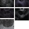
Sohini Sameera and Michel Kahaleh Rutgers Robert Wood Johnson Medical School, New Brunswick, NJ, USA Gastric outlet obstruction (GOO) is a common complication of gastrointestinal malignancies and inflammatory processes; it can lead to significant weight loss and nutritional deficiencies [1,2]. The current standard of care for treating GOO is surgical gastrojejunostomy; however, invasive intervention can be challenging in this patient population due to chronic malnourishment. The procedure itself carries a high complication rate that approaches more than 40% [1,2]. In addition, other adverse events associated with surgical bypass, such as delayed gastric emptying, prolonged hospital stays and overall increased cost, lead clinicians away from this treatment route. Endoscopic ultrasound‐guided gastrojejunal bypass (EUSGJ) has emerged as a minimally invasive alternative and may improve quality of life, especially in high‐risk surgical candidates. When comparing EUSGJ with surgical gastrojejunostomy, results regarding technical success tend to vary; one study found the technical success rate significantly higher in the surgical cohort while another study found no difference [3,4]. Furthermore, the rate of adverse events has been shown to be significantly lower in the EUSGJ cohort [4]. A meta‐analysis demonstrated the immediate safety and efficacy of EUSGJ as between 88 and 95%, with a pooled clinical success rate of 90% [5]. Finally, one study determined a significant difference between patients’ ability to eat before and after the EUSGJ, indicating great potential for clinical use [6]. Although there is a need for more data prior to endorsing EUSGJ as standard of care for GOO, the early results are promising and suggest that the procedure is safe and efficacious. EUSGJ was first introduced by Fritscher‐Ravens et al. in 2003; it has since been studied and various techniques have been proposed [7]. However, it is only when lumen‐apposing metal stents (LAMS) arrived that the procedure became more widespread [8]. Initially performed over a wire targeting a loop of bowel inflated with saline injection, the development of cautery‐enhanced LAMS (Hot AXIOS™, Boston Scientific, Natick, MA) made the procedure faster and easier to perform [2,8,9]. After identification of a loop of bowel appropriately distended by placement of a nasojejunal catheter or direct injection of contrast into the targeted lumen (Figure 47.1), a cautery‐enhanced LAMS is advanced through the stomach into the small bowel lumen. The first flange of the LAMS is then deployed, allowing the endoscopist to approximate the small bowel to the stomach (Figure 47.2) before deploying the second flange into the stomach (Figures 47.3 and 47.4). A commonly reported complication is stent misplacement. A case series examining international centers reported adverse events in 3 of 26 patients (11.5%) [8]. Two of these patients had stent misplacement, one of whom developed fulminant peritonitis. This patient, who had a long‐standing history of malignancy with peritoneal carcinomatosis, died the day after EUSGJ. In another case series by Itoi et al., 2 of 20 of patients (10%) experienced stent misplacement, which was quickly corrected with stent removal and conservative management [6]. Other less common complications include postprocedural pain, bleeding, and perforation. One patient in the Tyberg et al. study [8] experienced postprocedural pain and ultimately underwent a surgical gastrojejunostomy while another patient required a transfusion for failed LAMS placement without fistula closure. Figure 47.1 Puncture of the targeted bowel under endosonography. Source: Aloka. Figure 47.2 Deployment of the lumen‐apposing metal stent into the small bowel and connected to the stomach. Figure 47.3 Balloon dilatation of the lumen‐apposing metal stent. Figure 47.4 Visualization of the bowel through the fully distended lumen‐apposing metal stent.
47
How to do an EUS‐guided Gastrojejunostomy
Introduction
Technique (Video 47.1)
Complications


Related posts:


Stay updated, free articles. Join our Telegram channel

Full access? Get Clinical Tree


