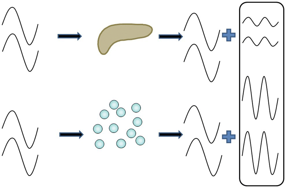
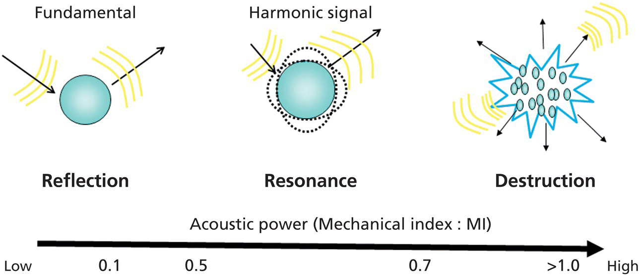
Yasunobu Yamashita and Masayuki Kitano Second Department of Internal Medicine, Wakayama Medical University, Wakayama, Japan Endoscopic ultrasonography (EUS) is considered one of the most reliable and efficient diagnostic modalities for digestive diseases, since it demonstrates better outcomes than other modalities with regard to spatial resolution. However, one of its primary limitations is differential diagnosis, which particularly focuses on vascularity to characterize digestive diseases. To address this issue, a number of studies have employed contrast‐enhanced harmonic EUS (CH‐EUS) to diagnose digestive diseases. Contrast harmonic imaging is based on the following principle: upon exposure to the ultrasound beams, microbubbles in the contrast agent either disrupt or resonate and subsequently release several harmonic signals. Exposure to ultrasound waves leads to the production of harmonic components in tissues and microbubbles that are integer multiples of the fundamental frequency; furthermore, microbubbles generate higher harmonic content than tissues. Selectively depicting the second harmonic component facilitates a better visualization of signals from microbubbles than those from the tissue (Figure 49.1). Therefore, this technology can detect signals without Doppler‐related artifacts from microbubbles in vessels that demonstrate extremely slow flow rates and is used to successfully characterize vascularity. Figure 49.2 demonstrates the impact of exposure to ultrasound beams on microbubble behavior, according to a mechanical index (MI). A dual screen with fundamental B‐mode and CH‐EUS images should be displayed. The CH‐EUS echo image should be dark enough to permit the visualization of the few signals from the tissue, before administering the contrast agent. The focal point should be set at the bottom of the target lesion. Before performing CH‐EUS, the target lesion should be imaged as close as possible, since ultrasound beam penetration via CH‐EUS is inferior to that with fundamental B‐mode. A bolus injection of contrast agent (Sonazoid, 15 ml/kg body weight; Sonovue, 2.4–4.8 ml per patient; Definity, 10 ml/kg body weight) is administered through a peripheral vein. Signals from the contrast agent appear after 10–15 seconds and peak at approximately 20 seconds (Figure 49.3). Characterizing a lesion involves classifying the enhancement patterns of the surrounding tissue into four patterns (avascular, hypovascular, isovascular, and hypervascular) (Figure 49.4). Additionally, enhancement patterns observed in the tumor are classified as either homogeneous (uniform enhancement of the entire tumor) or inhomogeneous enhancement (tumor enhanced at different levels) (Figure 49.5). Figure 49.1 Principle of contrast harmonic imaging. Figure 49.2 Behavior of microbubbles when exposed to ultrasound beams based on the mechanical index.
49
How to do Contrast‐enhanced EUS
Introduction
Principle of CH‐EUS
Critical points for performing CH‐EUS
Related posts:
 How to do EUS‐guided Radiofrequency Ablation of Pancreatic Neuroendocrine Tumors
 Biliary Tract Pathology
 How to Inject Chemotherapeutic Agents
 How to do EUS‐guided Treatment of Gastric Varices
 How to use ex vivo Models in Teaching Therapeutic Endoscopic Ultrasound
 How to Perform Endoscopic Ultrasound‐directed Transgastric Endoscopic Retrograde Cholangiopancreatography (EDGE)
Stay updated, free articles. Join our Telegram channel

Full access? Get Clinical Tree


