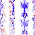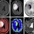


Through extensive technological advancements and numerous attempts, the first simultaneously acquired in vivo PET and MR images were demonstrated by Catana and colleagues in rodents. Subsequently, the human PET/MR scanner became available with an MR-compatible PET insert housed in a 3-T MR scanner. Since then, a plethora of publications reporting results on continuing technical development, comparisons with PET/CT, and initial clinical experience had been published by many groups. With the FDA approval, hybrid PET/MR is no longer a research tool but clinically applicable.
Since receiving FDA approval, clinical applications of PET/MR have grown rapidly. Nevertheless, PET/MR remains a relatively new and expensive technology and thus will benefit from continuing technical development. In particular, developing novel MR-based attenuation correction approaches is one of the most actively pursued areas. While convincing results using MR-based attenuation correction have been reported, additional developments for anatomic regions beyond the brain are needed (please see Yasheng Chen and Hongyu An’s article, “Attenuation Correction of PET/MR Imaging,” in this issue). Dr Lalush illustrates how PET image quality can be improved by incorporating anatomic information as well as motion information from MR. Drs Boada, Koesters, Block, and Chandarana further demonstrate how motion information derived from simultaneously acquired MR images enables a reduction of motion blur in cardiac exams and small nodule detection in the lungs.
Moving PET/MR into clinical practice is challenging and requires subspecialized radiologic expertise in both PET and MR imaging (please see the article by Samuel Galgano, Zachary Viets, Kathryn Fowler, et al, “Practical Considerations for Clinical PET/MRI,” in this issue) as well as trained technologists. The immediate clinical applications for PET/MR have been for disease sites where the high soft tissue contrast of MR is important and therefore clearly superior to CT. These sites include the brain (please see Michelle M. Miller-Thomas and Tammie L.S. Benzinger’s article), head and neck (please see Yueh Z. Lee, Joana Ramalho, and Brice Kessler’s article), bone marrow (please see Shetal N. Shah and Jorge D. Oldan’s article), and pelvis (please see Jorge D. Oldan, Shetal N. Shah, and Tracy Lynn Rose’s article). In addition, MR is being used increasingly for cardiac imaging, and the combination of cardiac MR imaging and specific PET biomarkers can be an increasingly powerful tool for cardiovascular disease (please see the article by Jeffrey M.C. Lau, Richard Laforest, Felix Nensa, et al). Finally, CT has been the mainstay for anatomically based radiation therapy treatment planning, but PET/CT and MR imaging are increasingly used to refine the treatment targets, enabling higher doses to be delivered to targeted areas within the tumor and reducing dose to normal tissues. PET/MR would be a natural extension to functionally guided radiation therapy (please see Tong Zhu, Shiva Das, and Terence Wong’s article). For all patients, but particularly those that are radiation sensitive such as the pediatric population, the elimination of the CT portion of the study with PET/MR can reduce the radiation dose to one-half or less compared with PET/CT (please see Yueh Z. Lee and Franz Wolfgang Hirsch’s article, “Pediatric Applications of Hybrid PET/MRI,” in this issue).
Currently, the vast majority of PET imaging is with FDG. However, the development and availability of new PET tracers are accelerating. As this occurs, we expect to rely more on MR imaging for accurate delineation of functional tumor (using diffusion-weighted imaging, for example), and selective PET tracers (for example, hypoxia, receptor, proliferative markers) to evaluate and target subsets of tumor cells that may be either susceptible or resistant to therapy. We expect a significant expansion in the clinical applications of PET/MR as these new metabolically selective PET tracers become available in the future.
The development of PET/MR has opened up many opportunities for improved clinical imaging and interesting challenges for the research community. This issue of Magnetic Resonance Imaging Clinics of North America provides a survey of the opportunities enabled by PET/MR and some of the current challenges and limitations.
Stay updated, free articles. Join our Telegram channel

Full access? Get Clinical Tree


