CHAPTER 1 Temporal Bone
Embryology
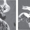
The temporal bones contain the sensory organs of hearing and balance, and these are located along the lateral and basal aspect of the skull. Each temporal bone consists of five parts: the squamous, the mastoid, the tympanic, zygomatic, and petrous segment (Fig. 1-1A). Important vascular channels, including the carotid artery and jugular vein, course through the temporal bone. The temporal bone is intimately related to the dura of the middle and posterior cranial fossa.
Squamous Portion
The squamous portion is composed of a flat plate of bone that forms the lateral wall of the middle cranial fossa and medial wall of the high masticator space. The outer surface is smooth and convex; it provides attachment to the temporalis muscle. A curved line (the temporal line) runs upward and backward along the posterior part, serving as an attachment for the temporalis fascia. The outer surface also demonstrates a vertical groove for the middle temporalartery. The squamous portion contributes to the posterior segment of the zygomatic arch and forms the anterior, or articular, portion of the mandibular fossa. The mandibular fossa is bounded anteriorly by the articular tubercle and posteriorly by the tympanic part of the temporal bone, which separates it from the external acoustic meatus.
The internal surface of the squamous temporal bone is concave and forms the lateral wall of the middle cranial fossa (Fig. 1-1B). It protects the lateral aspect of the temporal lobe of the brain. Branches of the middle meningeal vessels are related to the inner surface. Its superior border is formed by the squamosal suture, where it contacts the parietal bone. The anteroinferior border is thick and serrated; it articulates with the greater wing of the sphenoid.
Tympanic Portion
The tympanic portion is a U-shaped, curved bony plate that forms much of the external auditory canal (EAC) and the posterior (nonarticular) part of the glenoid fossa. It is located below the squamous portion and in front of the mastoid portion. The posterosuperior surface is concave, forming the anterior wall, the floor, and part of the posterior wall of the bony external acoustic meatus. The anteroinferior surface constitutes the posterior boundary of the mandibular fossa, and is related to the retromandibular part of the parotid gland.
The lateral border is free and gives attachment to the cartilaginous part of the external acoustic meatus. The tympanic sulcus is located on its medial aspect, providing the attachment for the tympanic membrane.
Styloid Portion
The styloid process is a thin and pointed bony projection that extends inferiorly from the posterolateral aspect of the inferior surface of the petrous bone. The proximal part is ensheathed by the vaginal process of the tympanic portion and it is located at the anterior margin of the stylomastoid foramen.
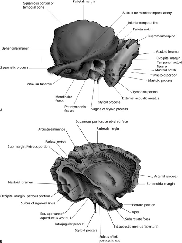
Figure 1-1
Related posts:

Stay updated, free articles. Join our Telegram channel

Full access? Get Clinical Tree


