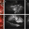
Fig. 5.1
Operative findings of metastatic parapharyngeal space from hypopharyngeal cancer
(a): Arrow represents a metastatic lymph node
(b): Dotted circle demonstrates the metastatic lymph node detected by ICG fluorescence Imaging over soft plate
(c): ICG fluorescence Imaging detects the metastatic lymph node (arrows)
5.3.4 Discussion
The handheld HEMS can significantly aid in the detection and visual confirmation of pharyngeal tumors located behind the oral cavity or nasal cavity by ICG-enhanced imaging with vivid color. Of note is that it can be freely operated intraoperatively by surgeons. Because of the high sensitivity of ICG fluorescence imaging, ICG fluorescence imaging under HEMS can assist surgeons in the identification and resection of tumors which have invaded the parapharyngeal space behind the internal carotid artery, internal jugular vein, and lower cranial nerves.
We have demonstrated a successful method of distinguishing cancer from healthy tissue and the optimum surgical time with HEMS in animal models [14]. Application of endoscopic and robotic surgery for the parapharyngeal space lesions enables surgeons to perform minimally invasive surgery with superior results [15]. However, we need to be able to detect parapharyngeal tumors in deeper and invisible areas when palpation is not possible. This is required in order to resect tumors safely and can be aided through effective tumor detection carried out with ICG fluorescent imaging.
5.3.5 Conclusion
ICG fluorescence imaging is effective for the detection and resection of the parapharyngeal space tumors which enables greater preservation of functions.
5.4 Lymphatic Chemotherapy for Head and Neck Cancer
According to the sentinel theory, metastatic lymph nodes are directly connected with primary tumors via lymphatic canals. Lymphatic chemotherapy is defined as chemotherapy using lymphatic canals between metastatic lymph nodes and primary tumors. This therapy is viewed as an ultimate cancer treatment which is both highly effective and noninvasive.
(a)
Clinical N0 Cases (Occult Neck Metastasis)
In the following, we consider a newly developed lymphatic chemotherapy procedure targeting the SLN using intra-arterial (I-A) chemotherapy for oral cancer in order to improve prognosis and to preserve organs while avoiding surgical complications [11, 16–18]. Our concept of lymphatic chemotherapy is shown in Fig. 5.2. Namely, the schema of lymphatic chemotherapy demonstrates an anticancer drug administered to the primary cancer which moves selectively to SLNs via lymphatic canals. As a result, the anticancer drug is accumulated in the SLNs and results in a higher anticancer drug concentration in the SLNs than non-SLNs. Because cis-diamminedichloroplatinum (CDDP) is the most promising drug for the treatment of head and neck cancers, we have adopted intra-arterial chemotherapy administered to the primary cancer so as to increase the CDDP concentration. To examine the potential advantages, we compared the CDDP concentration of SLNs with that of non-SLNs. The mean CDDP concentrations in the SLNs and non-SLNs were 1.2 μg/g and 0.35 μg/g, respectively. Our ICG fluorescence procedure revealed that all metastatic lymph nodes, including SLNs, were without false-negative SLNs. However, of 7 metastatic lymph nodes, one was not identified by means of the conventional radiocolloid method [11]. Detection of SLNs was clearly demonstrated by ICG fluorescence imaging (Fig. 5.3). The mean number of SLNs was 5.6 (3–8). ICG fluorescence imaging showed a greater number of SLNs than seen when injecting radiocolloid intratumor (mean 3.4). SLNs detected by ICG fluorescence imaging included all of the SLNs detected by the conventional radioactive method.
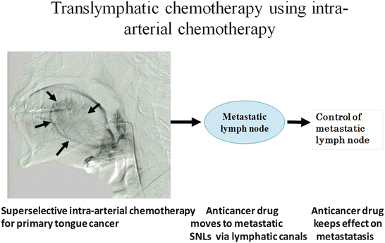
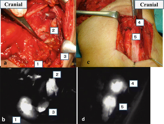
Fig. 5.2
Schema of lymphatic chemotherapy using intra-arterial chemotherapy
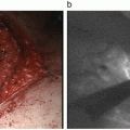
Fig. 5.3
Intraoperative navigation surgery using ICG fluorescence imaging
Upper figures (a, c) illustrate intraoperative navigation surgery by natural light; lower figures (b, d) illustrate intraoperative navigation surgery by ICG fluorescence imaging.
Number (1–5) means SLNs. (a) and (b) represent level II and III dissection. (c) and (d) represent level III and IV dissection
(b)
Clinical Neck Metastasis Cases
In the case of oral cancer with neck metastasis, modified radical neck dissection is often conducted to prevent cancer recurrence. However, this procedure can be the cause of severe postoperative complications. Instead of neck dissection and the associated surgical complications, minimally invasive lymphatic chemotherapy targeting neck metastasis based on the sentinel concept may contribute to the development of a revolutionary cancer treatment. This could potentially provide a superior method for the treatment of head and neck cancer which has been a key objective for researchers over many years. In our research, we consider I-A chemotherapy administered to primary oral cancers as not only organ preservation therapy but also a newly developed lymphatic chemotherapy targeting neck metastasis in order to improve prognosis.
In our examination we evaluate the effect of lymphatic chemotherapy targeting neck metastases in patients with oral cancer (T3N2bM0) by measuring CDDP concentrations in metastatic lymph nodes and pathological effects [19].
5.4.1 Methods
Seven patients with tongue cancer (cT3N2bM0) were treated by intra-arterial chemotherapy as neoadjuvant chemotherapy. After a week of chemotherapy, patients underwent surgical treatment. Intra-arterial chemotherapy was administered at 75 mg/m2 of CDDP two times weekly. At the beginning of surgery, 5 mg of ICG was administered to the lingual artery. SLNs were detected using ICG fluorescence imaging and a conventional radioactive method. The effect of lymphatic chemotherapy was evaluated based on the Ohboshi and Shimosato classification using the surgical specimens [20] and apoptosis using Trevigen’s apoptosis detection kit.
5.4.2 Results
The mean CDDP concentrations in the metastatic lymph nodes and non-SLNs were 2.35 μg/g and 1.08 μg/g, respectively (p = 0.034). Of 27 metastatic nodes, 24 (89 %) were identified by ICG fluorescence imaging; however, only 18 (67 %) were identified by the conventional method (p = 0.043). Of 22 measurable metastatic nodes, eight responded (partial response) and 14 did not respond (stable disease). Vast metastatic cancer was almost entirely diminished and resulted in scar tissue. Apoptosis was detected in all 27 metastatic lymph nodes and a pathological effect was achieved (Fig. 5.4).
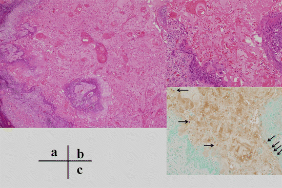
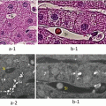
Fig. 5.4
Pathological findings of metastatic lymph nodes after chemotherapy
(a) Most of the metastatic cancer resulted in scar tissue; however, small cancer nests were residual (low-power magnification). (b) Cancer cells appeared to survive within small cancer nests (high-power magnification). (c) Many apoptotic bodies were detected within small cancer nests. Arrows indicate apoptotic cells
5.4.3 Discussion
We have recently demonstrated that intra-arterial chemotherapy for the treatment of primary tongue cancers is also effective as a means of lymphatic chemotherapy as it aids in the control of the subclinical metastatic tumors in SLNs (11). All SLNs were detected by ICG fluorescence imaging infused via the lingual artery in cT3N0M0 tongue cancer patients. However, of 27 metastatic lymph nodes, 24 (89 %) were detected by ICG fluorescence imaging infused via the lingual artery in seven cT3N2bM0 tongue cancer patients. The number of SLNs including metastatic lymph nodes resulting from I-A infusion was greater than that observed by means of a conventional injection into the tumor. Even in the case of micrometastatic SLNs, an afferent lymphatic canal was sometimes occluded by micrometastatic cancer based on sentinel navigation or CT lymphography [21]. In the current study, we also did not detect 9 (33 %) metastatic lymph nodes by conventional methods due to occlusion of afferent lymphatics from the tongue cancer (Fig. 5.5). However, the lymph nodes contained CDDP concentrations as high as 4.65 μg/g. This was because each lymph node has several afferent lymphatics and ICG or CDDP could move to metastatic lymph nodes via several other afferent lymphatics in the case of intra-arterial infusion. CDDP was released continuously from the primary tongue cancer via the lymphatic canal for a period of over more than 1 week. CDDP was selectively accumulated in metastatic lymph nodes and continued to affect metastatic lymph nodes over a long period.
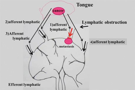
Fig. 5.5
Schema of increasing CDDP concentration of metastatic lymph nodes
Despite occlusion of afferent lymphatics from the tongue cancer, each lymph node has several afferent lymphatics, and ICG or CDDP could move to metastatic lymph nodes via several other afferent lymphatics in the case of intra-arterial infusion. (1) Occluded afferent lymphatics (red arrow). (2–4) Open afferent lymphatics (black arrows)
Our intra-arterial chemotherapy is expected to contribute not only to primary organ preservation but also to positive prognosis by controlling the metastatic SLNs. Preservation of patients’ quality of life in advanced cT3N2bM0 tongue cancer can be effectively achieved by intra-arterial chemotherapy and targeting metastatic lymph nodes with lymphatic chemotherapy.
As for ICG fluorescence imaging, metastatic SLNs were clearly detected even in close proximity to the primary tumor and “shine through” could be avoided. The ICG fluorescence imaging procedure demonstrated higher success rates for the detection of SLNs in patients with tumors located in the tongue than the radioactivity method.
5.4.4 Conclusion
CDDP concentrations in metastatic nodes were significantly higher than those in non-SLNs. This novel drug delivery system is feasible for lymphatic chemotherapy targeting metastatic nodes in patients with cT-3N2bM0 tongue cancer.
Related posts:
 ICG Videoangiography in Neurosurgical Procedures
 Blood Supply Visualization for Reconstruction During Esophagectomy
 Indocyanine Green Fluorescence-Navigated Sentinel Node Navigation Surgery (SNNS) for Cutaneous Malignant Melanoma and Extramammary Paget’s Disease
 Anatomical Hepatectomy Using Indocyanine Green Fluorescent Imaging and Needle-Guiding Technique
 Basic Aspects of ICG Fluorescence Imaging of the Liver
 Principle and Development of ICG Method
Stay updated, free articles. Join our Telegram channel

Full access? Get Clinical Tree


