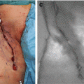
Fig. 24.1
Inflow to the liver. Soon after the injection of ICG, ICG fluorescence flow recognized in common hepatic artery (HA) and then in portal vein (PV). There was a several-second time lag between the fluorescent image of HA and PV. Pictures of both (a) and (b) were taken at the operation of pancreaticoduodenectomy. The luminal flow of hepatopetal portal vein was clearly identified with ICG fluorescence (c) which was shown in cirrhotic patient. LL left lobe
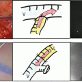
After we observed these flows, we saw fluorescence in the entire liver. The fluorescence spread homogeneously throughout the liver within several seconds (Fig. 24.2a–c). During a hepatectomy, the unilateral fluorescent images were seen after clamping the right or left vascular branch of the liver at the hepatic hilum. The tattooing of these hepatic segments was found to be very useful for hepatic surgeries. In addition, the Cantlie line was clearly recognized immediately after the injection of ICG (Fig. 24.2a and b). These findings were very informative for the evaluation of the fluorescence not only in the normal liver but also the cirrhotic liver and tumors in the liver.
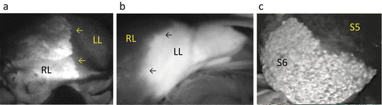
Fig. 24.2
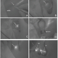
Early hepatic phase. The ICG fluorescence was seen soon after the injection of ICG. These pictures were taken during a hepatectomy ((a) left hepatectomy, (b) right hepatectomy, and (c) S6 segmentectomy). RL right lobe, LL left lobe, → Cantlie line. The cirrhotic changes are seen in panel (c)
The liver began to fluoresce at 40–50 s after the intravenous administration of ICG, which was taken into hepatocytes from the liver sinusoids. The fluorescence then reached a peak for several minutes. This peak was maintained for several hours. At approx. 30 min from the start of the ICG administration, ICG was discharged into the biliary tract via the bile canaliculi. These phenomena reflect the 15-min ICG load test value. The length of time from the uptake of ICG by hepatocytes until the ICG is discharged into the bile duct is relatively short, approx. 10–20 min.
24.2.2 Biliary Phase
One hour after a systemic injection of ICG, we observed the fluorescent images in the biliary tract, which can be described as cholangiography using ICG (Fig. 24.3a, b and c). In an ex vivo experiment, we were also able to detect the fluorescence of a mixture of ICG and bile juice (Fig. 24.3a and b). We evaluated the appropriate dilution ratio of bile juice and ICG solution. The maximum brightness of the ICG fluorescence was observed using the dilution of ICG solution to approx. 100–200-fold.
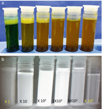
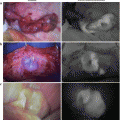
Fig. 24.3
Ex vivo finding of the fluorescence intensity of ICG changes with variations in the ICG concentration. (a) Before. (b) After excitation by LED: 1 ml of the original ICG solution and 10 ml bile juice combined: the original ICG solution: ICG 25 mg with 10 mL distilled water. The maximum brightness of the ICG fluorescence was observed using the dilution of ICG solution to approx. 100–200-fold. (c) Biliary phase. Fluorescent endoscopic picture at the operation of endoscopic cholecystectomy. Around 30 min after injection of ICG, the fluorescence biliary image was obtained, which is applied as the cholangiography at the operation of cholecystectomy
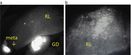
24.2.3 Late Hepatic Phase
ICG can be injected preoperatively as an ICG liver test to provide fluorescent images of liver tumors. We have evaluated the fluorescent images of the noncancerous liver at the late hepatic phase several days after an injection of ICG (Fig. 24.4a and b). In the normal liver, the fluorescence was not seen (Fig. 24.4a), but the retention of fluorescence was observed showing spotty or nodular patterns (Fig. 24.4b).
 ICG Fluorescent Image-Guided Surgery in Head and Neck Cancer
 Regional Lymph Node Dissection Assisted by Indocyanine Green Fluorescence Lymphography and Angiography for Stage III Melanoma
 Fluorescence Imaging for Intraoperative Identification of Pancreatic Leak
 Intraoperative Detection of Hepatocellular Carcinoma Using Indocyanine Green Fluorescence Imaging
 Indocyanine Green Fluorescent Lymphography and Microsurgical Lymphaticovenous Anastomosis
 Principle and Development of ICG Method


Related posts:
ICG Fluorescent Image-Guided Surgery in Head and Neck Cancer
Regional Lymph Node Dissection Assisted by Indocyanine Green Fluorescence Lymphography and Angiography for Stage III Melanoma
Fluorescence Imaging for Intraoperative Identification of Pancreatic Leak
Intraoperative Detection of Hepatocellular Carcinoma Using Indocyanine Green Fluorescence Imaging
Indocyanine Green Fluorescent Lymphography and Microsurgical Lymphaticovenous Anastomosis
Principle and Development of ICG Method
Stay updated, free articles. Join our Telegram channel
Full access? Get Clinical Tree
