Fig. 34.1
Structure and chemical reaction of glutaryl phenylalanine hydroxymethyl rhodamine green (gPhe-HMRG). gPhe-HMRG was used with a small amount of trypsin (chymotrypsin probe) to detect chymotrypsinogen in pancreatic juice leakage; gPhe-HMRG emits fluorescence when it is hydrolyzed by chymotrypsin
34.2 Pancreatic Chymotrypsin as a Target Substance for Fluorescence Imaging
When increased fluorescence intensity by the novel chymotrypsin probe was calculated in vitro by evaluating fluorescence intensity for the various chymotrypsin concentration samples, we confirmed the possibility of quantitative evaluation of chymotrypsin activity with sufficient linearity (Fig. 34.2). Yamashita et al. had previously revealed that this probe could specifically identify pancreatic juice in surgically drained fluids after pancreatic resection [10]. In the same study on patients undergoing pancreaticoduodenectomy, fluorescence imaging, after the chymotrypsin probe was sprayed on filter paper transcribing the pancreatic stump, enabled visualization of a pancreatic leak and accurate estimation of symptomatic postoperative PF [10].
Fig. 34.2
Relationship between the chymotrypsin concentration and the increasing rate of fluorescence intensity. The rate of fluorescence intensity increased with an increase in the concentration of chymotrypsin. We confirmed that the chymotrypsin probe could quantitatively detect the activity of chymotrypsin with sufficient linearity
It is also an innovation that the chymotrypsin probe targets the type of protease—not amylase—that can potentially cause severe tissue damage and lead to serious postoperative complications associated with PFs. Postoperative PF is presently diagnosed by evaluating amylase levels [13, 14] using International Study Group on Pancreatic Fistula classification grades [15]. It is unlikely that amylase, which is simply a glycolytic enzyme, directly triggers the complications stemming from postoperative PFs. Some authors have indicated that the current definition of postoperative PF, which is based on amylase levels in drainage fluids, does not always reflect complication severity [16, 17]. Pancreatic protease secretion can result from several factors, such as diet, pancreatic disease, and stimulatory or inhibitory mediators, and may not parallel amylase secretion [18, 19].
34.3 Application of the Chymotrypsin Probe for Pancreatic Resection in a Swine Model
Ideally, the chymotrypsin probe should be sprayed directly onto the pancreatic stump in the patient’s abdominal cavity to enable surgeons to accurately suture the pancreatic leak location. However, such usage has not yet been approved in humans (pending completion of safety trials); therefore, this technique was simulated using a swine model. Chymotrypsin probe was sprayed on the pancreatic stump in abdominal cavity, following full mobilization and division of the pancreatic parenchyma with the Focus Ultracision Harmonic Scalpel (Ethicon Endo-Surgery Inc., Cincinnati, OH, USA). Five minutes after the chymotrypsin probe was sprayed onto the pancreatic stump, fluorescence imaging using a FluorVivo imaging system (INDEC, Inc., Santa Clara, CA) with a blue-filter setting (excitation, 470–490 nm; emission, 515 nm long pass) enabled identification of pancreatic juice leaking point (Fig. 34.3). Fluorescence of pancreatic juice was also identifiable by naked eye examination through light-blocking glasses (515 nm long pass), which enabled surgeons to apply pinpoint sutures and confirm pancreatic leak arrest. This technique may also enable surgeons to determine whether prophylactic abdominal drainage during pancreatic resection is needed, based on protease activities of pancreatic juice in each patient, as well to determine the presence or absence of pancreatic leak after closure/reconstruction of the pancreatic remnant.
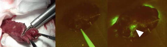
Fig. 34.3
Fluorescence images of pancreatic juice leaking from a pancreatic stump using the chymotrypsin probe in a swine model. After the division of the pancreatic parenchyma with the Harmonic Scalpel (left), chymotrypsin probe was sprayed on the pancreatic stump in abdominal cavity (middle). Five minutes after the chymotrypsin probe was sprayed onto the pancreatic stump, fluorescence imaging enabled identification of pancreatic juice leaking point (right, arrowhead)
34.4 Conclusion
In this chapter, mechanistic background and potential roles of fluorescence imaging using chymotrypsin probe are demonstrated. Following preclinical safety trials, first-in human study will be conducted in order to evaluate efficacy of intraoperative real-time fluorescence imaging of pancreatic leak in reducing incidence and severity of postoperative PF.
Acknowledgment
Related posts:
 ICG Fluorescent Image-Guided Surgery in Head and Neck Cancer
 Regional Lymph Node Dissection Assisted by Indocyanine Green Fluorescence Lymphography and Angiography for Stage III Melanoma
 Indocyanine Green Fluorescence-Navigated Sentinel Node Navigation Surgery (SNNS) for Cutaneous Malignant Melanoma and Extramammary Paget’s Disease
 Anatomical Hepatectomy Using Indocyanine Green Fluorescent Imaging and Needle-Guiding Technique
 Basic Aspects of ICG Fluorescence Imaging of the Liver
 Principle and Development of ICG Method
Stay updated, free articles. Join our Telegram channel

Full access? Get Clinical Tree


