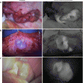
Fig. 8.1
Advantages of NIR fluorescence over visible light: Tissue penetration (upper panel): lymphatic vessels under the skin and lymph nodes in the fatty tissue can be detected by fluorescence. High sensitivity (lower panel): lymph nodes stained by ICG can be judged with high sensitivity
8.2 Principle
8.2.1 Molecular Fluorescence of ICG
Molecular fluorescence of ICG consists of a two-step process, excitation and emission (Fig. 8.2). When light at 765 nm is emitted at ICG molecules, they move from ground state to excited state (excitation). When the molecules return to ground state, a part of the absorbed energy is radiated as fluorescent emission at 830 nm. The fluorescent emission is at a longer wavelength than the fluorescent excitation. Since the combination of these wavelengths is specific to the ICG molecule, highly sensitive measurement is possible by selecting proper wavelengths in the light source and the detector.
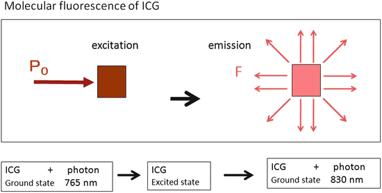
Fig. 8.2
Molecular fluorescence of indocyanine green (ICG): The wavelengths of fluorescent excitation and emission are specific to ICG. High signal-to-noise ratio can be obtained by fluorescence measurement. P 0 excitation light, F fluorescent light
The general law of fluorescence spectroscopy is shown in the equation below [4]. The quantum efficiency shows that only 1.2 % of absorbed energy is emitted as fluorescence. The equation also indicates the nonlinear relationship between the fluorescence intensity and the ICG concentration, meaning that quantitative measurement is difficult. When the concentration of ICG is greater than 0.08 g/l, the fluorescence intensity becomes weaker (quenching effect). However, the quenching effect usually does not disturb the lymphatic detection in the clinical setting, since the administered ICG is adequately diluted by the lymphatic fluid.
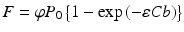
F: Intensity of fluorescent light
P0: Intensity of excitation light
φ: Quantum efficiency (ICG: 0.012)
exp: Natural log
ε: Absorption coefficient
C: ICG concentration
b: Cell size
8.2.2 Optical Properties of ICG in the Living Tissue
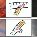
ICG has characteristic fluorescence spectra in the NIR wavelengths [5] (Fig. 8.3), ranging from 700 to 900 nm, which is called “an optical window.” This is advantageous to clinical application, because the NIR light can penetrate deep into the tissue without being absorbed by hemoglobin or water. However, when applied to the living tissue, light scattering becomes an important issue [6] (Fig. 8.4). The fluorescent excitation and emission are attenuated by scattering when the light travels in the tissue. Fat droplets in the axilla are the main scatterers [7]. In the preliminary experiment, an ICG fluorescent signal at the depth of 1 cm in the breast phantom was detectable, but detection was difficult as the depth increases. The limit of detectable depth by fluorescence in the axilla is supposed to be 1–2 cm.
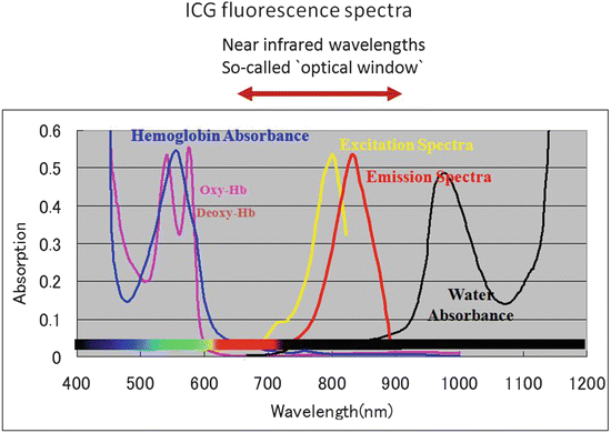
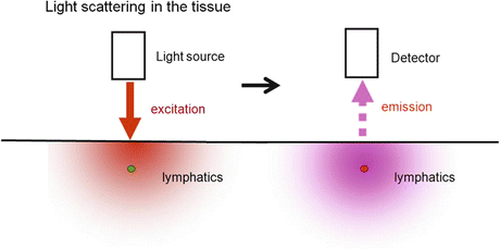
Fig. 8.3
Indocyanine green’s fluorescence spectra in the near-infrared wavelengths: The wavelengths between 700 and 900 nm are called the “optical window,” since the light at these wavelengths can penetrate deep into the tissue as escaped from the absorption by hemoglobin (Hb) and water
Fig. 8.4
Attenuation of fluorescence by scattering in living tissue: The fluorescent light is attenuated by scattering while traveling in the subcutaneous tissue. A main scatterer is subcutaneous fat droplets
8.3 Clinical Development for Sentinel Node Biopsy in Breast Cancer
8.3.1 Equipment
An infrared fluorescence imaging system (photodynamic eye, Hamamatsu Photonics, Japan), which consists of light-emitting diodes at 760 nm as a light source, and a charge-coupled device (CCD) camera with a cut filter below 820 nm as a detector were used to measure NIR fluorescence images [8] (Fig. 8.5).
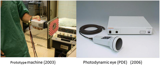
Fig. 8.5
Infrared fluorescence imaging system (Photodynamic eye, Hamamatsu Photonics, Japan): Prototype machine (left) and commercially available photodynamic eye (right)
8.3.2 Surgical Procedures
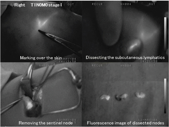
After induction of general anesthesia and sterilization of the operation site, 25 mg/5 ml indocyanine green (Diagnogreen 0.5 %; Daiichi Pharmaceutical, Tokyo, Japan) was injected in the subareolar breast tissue. Following a few seconds of massage, subcutaneous lymphatic drainage was then observed with fluorescent images (Fig. 8.6). Subcutaneous lymphatic streams could be detected over the skin usually within 1 or 2 min, and they disappeared before entering the axillary space. The subcutaneous lymphatic streams were marked again over the skin, and a 4-cm skin incision was made at the point where the lymphatic channels drained into the axilla. After skin incision, the subcutaneous lymphatics were more clearly observed. Green or fluorescent lymphatic channels were carefully dissected and traced until the first-drained lymph node was found in the axilla. The lymph node was then dissected with the surrounding fatty tissue. Usually, two or more lymph nodes embedded in the surrounding tissue were found. Fluorescence imaging was useful for the navigation of dissection. The lymphatic channels and nodes receiving ICG appeared as shining streams and spots with fluorescence in the fluorescence image. After removal, it was ensured that no fluorescent spot was left in the axilla. Lymph nodes in the dissected specimen were isolated from the surrounding fatty tissue, and we investigated whether each lymph node was fluorescent inside. All fluorescent lymph nodes were regarded as sentinel nodes.
 ICG Fluorescent Image-Guided Surgery in Head and Neck Cancer
 Regional Lymph Node Dissection Assisted by Indocyanine Green Fluorescence Lymphography and Angiography for Stage III Melanoma
 Fluorescence Imaging for Intraoperative Identification of Pancreatic Leak
 Intraoperative Detection of Hepatocellular Carcinoma Using Indocyanine Green Fluorescence Imaging
 Indocyanine Green Fluorescent Lymphography and Microsurgical Lymphaticovenous Anastomosis
 Detection of Hepatic Micrometastases from Pancreatic Cancer


Related posts:
ICG Fluorescent Image-Guided Surgery in Head and Neck Cancer
Regional Lymph Node Dissection Assisted by Indocyanine Green Fluorescence Lymphography and Angiography for Stage III Melanoma
Fluorescence Imaging for Intraoperative Identification of Pancreatic Leak
Intraoperative Detection of Hepatocellular Carcinoma Using Indocyanine Green Fluorescence Imaging
Indocyanine Green Fluorescent Lymphography and Microsurgical Lymphaticovenous Anastomosis
Detection of Hepatic Micrometastases from Pancreatic Cancer
Stay updated, free articles. Join our Telegram channel
Full access? Get Clinical Tree
