Fig. 17.1
Above: The design for en-bloc excision in Stage III melanoma case. The primary site is distal thigh. Clinical metastatic node was involved in regional groin nodal basin. Before en-bloc excision, the lymph vessels running through subcutaneous tissue were detected by ICG fluorescence lymphography and marked by black pen. Middle: A view of lymph vessels through PDE (Photodynamic Eye®, PDE; Hamamatsu Photonics, Hamamatsu, Japan) after elevation of skin flap. Below: Intraoperative view of en-bloc excision of lymph vessels and primary lesion (Figure 17.1 were previously published in leaflet for PDE and reprinted in this chapter with permission of Hamamatsu Photonics, Hamamatsu, Japan)
Before en-bloc excision, the ICG was injected evenly around a tumor lesion at a distance of 5 mm from the primary tumor margin. The ICG is dissolved at a concentration of 2.5 mg/mL in aqueous solution. 0.1 mL was injected at each of the four sites and the total dosage was 0.4 mL (ICG 1 mg). Lymph vessels, running from primary lesion into nodal basin, are detected by a near-infrared camera system (Photodynamic Eye®, PDE; Hamamatsu Photonics, Hamamatsu, Japan).
17.3 Decision of the Levels of Dissection of Regional Lymph Basin
For planning lymph node dissection, detection of lymph flow entering into lymph basin is important for decision of the excised levels in lymph basin. The axilla basin consists of three levels divided by edges of pectoralis minor muscle. In the case that primary lesion is located in upper extremity, lymph flow from primary lesion is usually entering level I of axilla, and dissection area includes levels I and II. If primary lesion is located near the shoulder, the additional lymph flow may run over pectoralis major muscle into level III (Fig. 17.2). The information of lymphography may help to decide whether the level III should be excised or not.
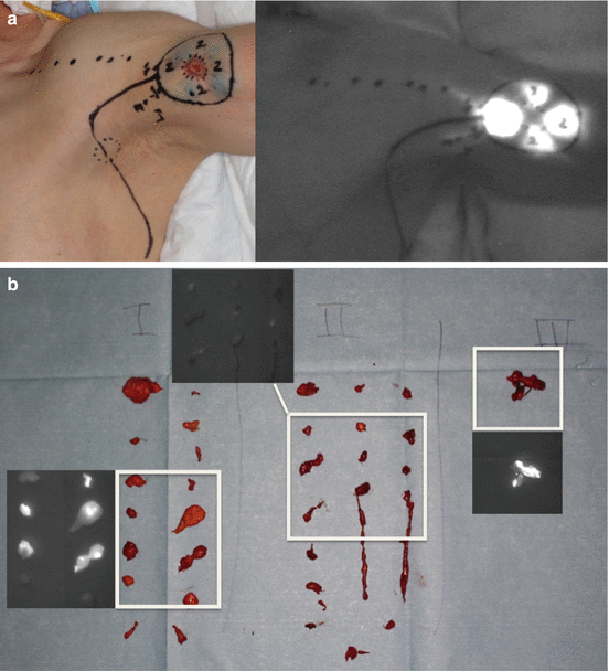
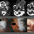
Fig. 17.2
 ICG Fluorescent Image-Guided Surgery in Head and Neck Cancer
 Blood Supply Visualization for Reconstruction During Esophagectomy
 Indocyanine Green Fluorescence-Navigated Sentinel Node Navigation Surgery (SNNS) for Cutaneous Malignant Melanoma and Extramammary Paget’s Disease
 Anatomical Hepatectomy Using Indocyanine Green Fluorescent Imaging and Needle-Guiding Technique
 Basic Aspects of ICG Fluorescence Imaging of the Liver
 Principle and Development of ICG Method


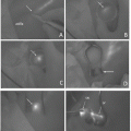
(a) Left: The design for axilla dissection in Stage III melanoma. Primary lesion located in proximal arm. Clinical metastatic node was involved in the axilla basin. Before dissection, ICG was injected intradermally around the primary site. Right: Same view as left photo through PDE. The lymph vessels running on pectoralis major muscle were detected and marked by black pen. (b) After dissection of levels I, II, and III, the lymph nodes were enucleated from dissected tissue at back table and divided into three levels. Small flames are view through PDE. Lymph nodes of levels I and III had fluorescence and level II did not. This confirmed the two lymph flows entering the axilla basin from primary tumor, one is into level I and the other is directly into level III (Figure 17.2 were previously published in leaflet for PDE and reprinted in this chapter with permission of Hamamatsu Photonics, Hamamatsu, Japan)
Related posts:
ICG Fluorescent Image-Guided Surgery in Head and Neck Cancer
Blood Supply Visualization for Reconstruction During Esophagectomy
Indocyanine Green Fluorescence-Navigated Sentinel Node Navigation Surgery (SNNS) for Cutaneous Malignant Melanoma and Extramammary Paget’s Disease
Anatomical Hepatectomy Using Indocyanine Green Fluorescent Imaging and Needle-Guiding Technique
Basic Aspects of ICG Fluorescence Imaging of the Liver
Principle and Development of ICG Method
Stay updated, free articles. Join our Telegram channel
Full access? Get Clinical Tree
