Fig. 40.1
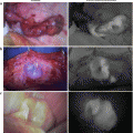
Comparison of lymphoscintigraphy with ICG lymphography
Several patterns of dermal backflow can be recognized in ICG lymphography (b), but in lymphoscintigraphy no such pattern is observed (a). Each of the lymphatic vessels in the artero-medial bundle can be recognized in ICG lymphography (c), but in lymphoscintigraphy, only the artero-medial bundle can be observed (a)
ICG has primarily been used as tracer for angiography. However, in 2007, Unno et al. introduced ICG-LG [11]. By using indocyanine green injection and a portable and easy-to-use infrared camera, real-time imaging of the lymphatic system was obtained. ICG is intradermally injected into the web space between the fingers, binds to albumin, and drains into the lymphatic vessels. The albumin-bound ICG is excited using a 760-nm infrared ray and emits an 840-nm infrared ray. The fluorescence of ICG in near-infrared wavelengths can deeply penetrate living tissue and is advantageous for obtaining noninvasive visual information about the lymphatic systems.
ICG-LG has been studied as not only a real-time imaging technique but also a method of diagnosing lymphedema. In 2008, Unno et al. measured the velocity of lymphatic flow with ICG-LG and reported that the decubitus position, exercise loading, and massage promoted lymphatic flow [13]. In 2011, Yamamoto et al. reported three types of dermal backflow (DB) patterns seen in lymphedema patients with ICG-LG: splash, stardust, and diffusion; they also reported relationships between the distribution of these DB patterns and clinical staging [14]. In 2012, Akita et al. reported the possibility that the splash type can serve as an early sign of lymphedema [15]. Nowadays, ICG-LG is gradually gaining acceptance as a screening examination for early-stage lymphedema.
40.3 Surgical Operations for Lymphedema and ICG Lymphography
Various surgical operations for lymphedema have been performed. There are mainly two categories of surgical operations: volume-reduction methods and reconstructions of lymph flow. Volume-reduction methods such as the Kondoleon procedure [16], Charles procedure [17], and liposuction [18] simply attempt to ablate the offending tissue, mainly fatty tissue. Reconstruction of lymph flow attempts to augment lymph flow or egress from the lymphedematous extremity by establishing communication between the congested lymphatic system and a competent drainage system such as deep lymphatic vessels or venulae. There are some classical reconstructions of lymph flow like the Degni procedure [19] and O’Brien [20] procedure, which involve anastomosis to the saphenous vein, but nowadays the mainstream procedure is lymphaticovenous anastomosis (LVA), which involves anastomosis to a venula [21]. All reconstructions of lymph flow first need to discover the thin, transparent lymphatic vessels within the adipose tissues. ICG-LG visualizes the lymphatic vessels clearly in real time without using radiation, resulting in an improved discovery rate of lymphatic vessels.
40.4 Protocol of ICG-LG
ICG lymphography appeared in the 2000s and is a relatively new method. Therefore, the protocol of ICG-LG has not been fully established in detail. Typical methods are shown.
40.4.1 Preparation of Examination
40.4.1.1 Examination Reagent
ICG has been produced as Diagnogreen® for injection (Daiichi Sankyo Company, Ltd., Tokyo). First, 25 mg of Diagnogreen powder is dissolved in 2 ml pure water. Second, the solution of Diagnogreen is added to 2 ml of 1 % Xylocaine® injection. The addition of Xylocaine relieves the pain of the injection of ICG solution. However, it is important not to add Xylocaine to the vial of Diagnogreen first because it cannot dissolve the powder; only water (2–10 ml) can.
40.4.1.2 Pain Control
As the pain of the injection of ICG is extreme, controlling the pain is important. Not only the addition of Xylocaine in solution but also the application of EMLA® cream is effective at reducing pain. Intradermal injection is more painful than subcutaneous injection.
40.4.1.3 Setup of Examination Room
The examination needs to be conducted in a dark room. Even the glimmer of a liquid crystal screen or infrared heater will disturb the observation of the deep lymphatic systems.
40.4.2 Ladder of Examination and Operation
40.4.2.1 Injections
Injections of ICG are performed in the intradermal or subcutaneous layer. It is necessary to consider the differences in lymphatic velocity and lymphatic uptake in ICG-LG.
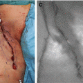
Our group injects both layers with 30-gauge needle: the intradermal layer receives 0.04 ml and subcutaneous layer receives 0.06 ml for a total of 0.1 ml for one injection point. In the lower extremity, ICG is injected into three points (Fig. 40.2a). In the upper extremity, ICG is injected into five points (Fig. 40.2b and c).
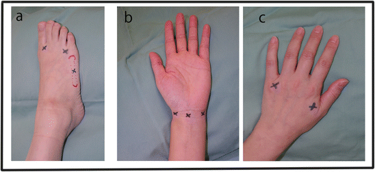
Fig. 40.2
Injection points of extremities in ICG lymphography
In the lower extremity, ICG is injected into three points: the first and fourth web space and the middle of the fifth metatarsal (a). In the upper extremity, the palm side is injected at the radial, the ulnar, and the middle of the wrist (b), and the dorsal side is injected at the first and fourth web space (c)
This method does not represent all of subcutaneous lymphatic systems because ICG is injected from the peripheral side. Therefore, it is necessary to consider how to inject ICG further.
40.4.2.2 Observations and Selection of Lymphatic Vessels for LVA
One of the most important functions of preoperative ICG-LG is to detect lymphatic vessels without fibrosis, but with high flow for anastomosis. Sometimes the point where dermal backflow (DB) originates can be observed. If there is no DB, it is very easy to detect good lymphatic vessels by ICG-LG. But if DB widespread, the lymphatic vessels will be hidden behind it. It is important to note that physical activity during the examination makes the lymphatic flow increase and, often, the DB area to spread (Fig. 40.3). Therefore, our group observes patients at rest. If lymphatic vessels cannot be observed, a soft massage to the puncture area is added. Lymphatic vessels are mapped as soon as they are observed.
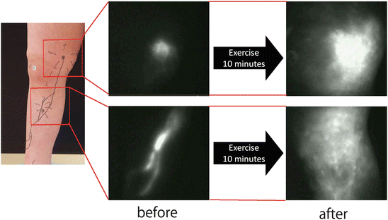
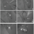
Fig. 40.3
Effects of exercise in ICG lymphography
Before exercise, lymphatic vessels can be observed clearly. But exercise makes dermal backflow appear and become widespread, hiding the lymphatic vessels
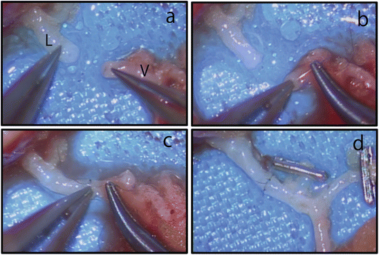
40.4.3 Skin Incision, Preparation, and Anastomosis of Lymphatic Vessels
Small skin incisions are performed above the lymphatic vessel. Most of the lymphatic vessels observed by PDE camera are located under the superficial fascia. Microsurgical lymphovenous anastomoses are performed between lymphatic vessels and venulae (Fig. 40.4). The most commonly performed techniques are direct end-to-end, end-to-side, or side-to-end microsurgical anastomoses. After anastomosis, a PDE camera can check the patency and lymphatic flow in the venula (Fig. 40.5).
 ICG Fluorescent Image-Guided Surgery in Head and Neck Cancer
 Regional Lymph Node Dissection Assisted by Indocyanine Green Fluorescence Lymphography and Angiography for Stage III Melanoma
 Fluorescence Imaging for Intraoperative Identification of Pancreatic Leak
 Intraoperative Detection of Hepatocellular Carcinoma Using Indocyanine Green Fluorescence Imaging
 Basic Aspects of ICG Fluorescence Imaging of the Liver
 Principle and Development of ICG Method


Related posts:
ICG Fluorescent Image-Guided Surgery in Head and Neck Cancer
Regional Lymph Node Dissection Assisted by Indocyanine Green Fluorescence Lymphography and Angiography for Stage III Melanoma
Fluorescence Imaging for Intraoperative Identification of Pancreatic Leak
Intraoperative Detection of Hepatocellular Carcinoma Using Indocyanine Green Fluorescence Imaging
Basic Aspects of ICG Fluorescence Imaging of the Liver
Principle and Development of ICG Method
Stay updated, free articles. Join our Telegram channel
Full access? Get Clinical Tree
