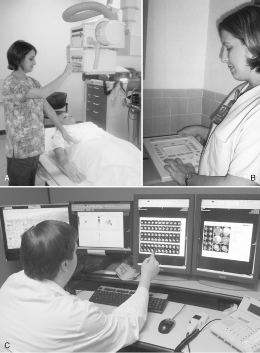
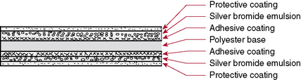
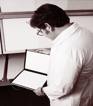
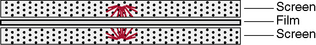
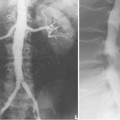
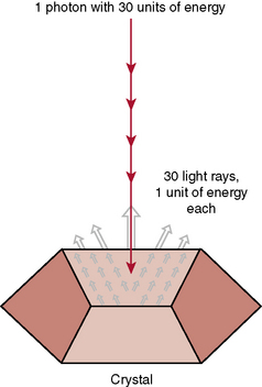
Imaging: Conventional and Digital

CHAPTER 10


Imaging


Conventional and Digital


Conventional and digital imaging


This chapter discusses several factors that affect the production of a radiographic image and several ways to produce the image. It includes discussions on analog or film images, as well as x-ray digital images. In digital radiography, the image is made in discrete units; whereas a film image is continuous. For every concept briefly discussed in this text, a substantial amount of theory and practical application will be presented in the classroom and in your clinical experience.


Conception in the Mind of the Physician


Once the physician has determined the need for and orders radiographic studies, the radiologic technologist is responsible for obtaining the best possible diagnostic images. The radiologic technologist evaluates the orders from the physician and greets the patient in the imaging department. Establishing a cordial relationship with the patient helps the radiologic technologist obtain the needed diagnostic test.


The diagnostic test begins with the positioning of the patient (Fig. 10-1, A). Next, the technologist determines the appropriate exposure factors to be used to place the image on the image receptor (Fig. 10-1, B). The image receptor may be the conventional film-screen or, most likely, a photosensitive phosphor plate as in computed radiography (CR) or a charged electronic device as in digital radiography, also known as direct digital radiography, (DR). The image receptor in CR is a photostimulable phosphor plate. The latent image is created when x-rays pass through the body part and strike the phosphor, some giving off light; however, some are held within the phosphor particles, creating a latent image. The image receptor in DR is a charged electronic device. The x-ray photons strike a scintillator or a photoconductor. The latent image is in the form of an electrical signal. In any case the image is latent; it is not visible to the human eye and needs to undergo processing to make it visible.


Regardless of the image receptor used, the anatomic part to be radiographed will be positioned and evaluated to determine the thickness of the part and the overall tissue density. In addition, when the image is made visible, it must be checked for diagnostic quality (Fig. 10-1, C). The technologist may then consult a technique chart for the proper exposure factors. The exposure factors that affect image quality are discussed later in this chapter.


Film-Screen Receptor


Radiographic film is composed of emulsion spread on a thin transparent sheet of polyester plastic. Except for special uses such as mammography, the emulsion is spread on both sides of the polyester base sheet. The emulsion is the image component of the film and consists of microscopic silver bromide crystals in a gelatin suspension (Fig. 10-2). The film is loaded in the cassette and sandwiched between the two screens (Fig. 10-3). The intensifying screens are made of crystals that will fluoresce when struck by x-rays (Fig. 10-4). The light emitted from the screen crystals will fluoresce when struck by x-rays. The light emitted from the screen crystals exposes the film (Fig. 10-5). Only approximately 5% of the radiographic film density is a result of the x-rays; 95% of the density is a result of light from the screen crystals.


To make the image visible, the film must be processed in a chemical solution, which is called the developer. The developer chemicals cause the crystals in the film that were struck by x-rays to become black metallic silver, which causes parts of the film to be darkened in places, creating the visible image.


Digital Radiography


DR was developed after CR and has evolved into several image-capturing devices using different elements to interact with x-rays. The elements used are cesium iodide, gadolinium oxysulfide, and selenium. The latent image in DR is recorded on the electronic device. Several methods are available for creating a visible image in DR. However, all require digital electronics to create a visible image.


DR can be produced by both direct and indirect methods. The indirect method is one in which the x-rays are first converted to light. Light is then emitted when x-rays strike a sensitive phosphor that then must be converted to an electric signal. Computers require an electric signal to generate a visible image. In direct digital imaging, x-rays interact directly with the element selenium, creating an electric charge. Direct digital imaging does not require a light-emitting phosphor. A computer program is essential to construct the image in all digital imaging. The advances in the development of computer technology account for the rapid growth in the use of this technology in medical imaging.


Advantages of Digital Radiography


Digital imaging has several advantages over conventional radiography. Producing an image without the use of film and without wet processing is one advantage. This time-consuming step is eliminated. Because a digital image is produced within a matter of seconds, the waiting time is shortened and the patient can remain on the examining table while the image is checked for diagnostic quality.


Even with faulty technique, the image can be manipulated and adjusted by the technologist and brought within an acceptable diagnostic range—a second important advantage. This capability eliminates retakes because the image can be altered without further exposure to the patient and is obviously a great convenience; consequently, efficiency in caring for the patient is improved. A word of caution is required here. Being able to manipulate the image is a mixed blessing. Some technologists who are careless with the exposure technical factors may over expose, knowing the image can be adjusted, giving the patient more radiation than is necessary. This troubling aspect was pointed out in research performed by Dr. Teri Fauber who studied the effects of insufficient and excessive radiation exposures. She concluded that the “study confirmed that extreme variability in radiation exposure will produce a diagnostic-quality digital image,” but she continues to remind operators that “over exposing patients is an objectionable practice” (Fauber, 2009). Digital imaging will tolerate a wide range of error in exposure technique, but proper technical factors are as important as ever in protecting the patient from unwanted or unnecessary radiation.


Objectionable fog, as is present in screen-film radiography, greatly minimized in digital imaging. This very important advantage results in an image with more contrast, thus enhancing the diagnostic quality of the image.


Storing images for future review is a significant advantage to be considered. In conventional radiography, the image is on film that must be labeled, stored, and filed for retrieval for future use. These necessities require a considerable amount of labor and storage space. In DR the image can be stored on a magnetic disk and on computer hardware and servers. Data stored in digital form can be retrieved for review at a later date and can be sent to distant locations for viewing and interpretation. In light of these advantages, the reasons why DR is so widely accepted and used are obvious.


You will learn more about digital imaging as you progress in your radiography program. Although the film-screen system has been around for over 100 years and is still the gold standard as far as resolution of detail is concerned, new technology is rapidly replacing it; consequently, some believe film radiographic imaging will be completely eliminated.


Factors affecting the image


The previous discussion concerned the image receptors, conventional film, phosphor plates as in CR, or electronic receptors as in DR. However, factors that influence the amount and energy of x-rays after passing through the patient are the same, regardless of the image receptor. In addition, the geometric factors are the same, and an understanding of these basic factors is essential to generating a diagnostic-quality image. The exceptions to some of the basic factors include two important points: (1) When the latent images are digitized to be made visible, density and contrast can be manipulated, within a limited degree, to a desired and acceptable image. (2) Detail in digital imaging is built in and inherent in the computer construction.


In screen-film radiographs, detail is measured in line pairs per millimeter. On film, 10 lines can be recorded with optimal technique. Digitized images do not record detail to this degree; however, because contrast is significantly better in digitized images, the recorded detail is easier to identify. The digital images actually “appear” to have more detail because of the acuity of the human eye.


The purpose of this text is not to examine in detail the physics involved in the production of x-rays but to provide an overview that will be helpful in the discussion of technical imaging factors. However, before discussing the factors that affect the image, the control panel of a radiographic machine should be examined to understand what controls are available for setting up a technique for obtaining optimal diagnostic quality.


With the typical radiographic machine, the controls to be adjusted are (1) time, (2) voltage, and (3) amperage. Because x-rays are not produced unless the voltage is very high (i.e., thousands of volts), the term kilovolt is used. However, with the very high voltage, low amperage must be used, and thus we usually speak in terms of milliamperage

Stay updated, free articles. Join our Telegram channel

Mar 2, 2016 | Posted by in GENERAL RADIOLOGY | Comments Off on Imaging: Conventional and Digital

Full access? Get Clinical Tree

Get Clinical Tree app for offline access