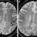
Infections of the head and neck vary in their clinical course and outcome because of the diversity of organs and anatomic compartments involved. Imaging plays a central role in delineating the anatomic extent of the disease process, identifying the infection source, and detecting complications. The utility of imaging to differentiate between a solid phlegmonous mass and an abscess cannot be overemphasized. This review briefly describes and pictorially illustrates the typical imaging findings of some important head and neck infections, such as malignant otitis externa, otomastoiditis bacterial and fungal sinusitis, orbital cellulitis, sialadenitis, cervical lymphadenitis, and deep neck space infections.
- •
Imaging plays a central role in delineating anatomic extent of infection, detecting complications, assisting in accurate diagnosis, and guiding drainage procedures.
- •
Computed tomography is the mainstay investigation in the imaging workup for head and neck infections.
- •
Magnetic resonance imaging is reserved for assessing intracranial extension and intramedullary bone marrow signal, and to monitor response to therapy.
- •
Ultrasound is preferred for assessment of lymph nodes, salivary glands, and neck abscesses, and plays an important role in imaging the pediatric population.
- •
Cross-sectional imaging is preferred in presurgical anatomic localization, particularly for deep-seated and locally extensive lesions.
Stay updated, free articles. Join our Telegram channel

Full access? Get Clinical Tree


