Urethral Diverticulum
Etiology
A urethral diverticulum is a focal outpouching of urethral tissue into the urethrovaginal space. It is thought to be due to postinflammatory dilatation and rupture of the periurethral glands (of Skene) into the urethra. Most urethral diverticula are acquired and occur in women between their third and sixth decades in age. The estimated prevalence of urethral diverticula is 0.6% to 6% of adult women. Congenital urethral diverticula are rare but have been reported in neonates and children.
Pathophysiology
It is widely believed that most diverticula result from infection of the periurethral glands. Other causes include urethral trauma from vaginal childbirth, instrumentation, and surgery.
Clinical Presentation
Women with urethral diverticula manifest a range of symptoms, including dysuria, increased urinary frequency, dyspareunia, and postvoid dribbling. Approximately 20% of patients are asymptomatic. Physical examination can reveal periurethral “bogginess,” a palpable cyst, and expression of mucus or pus through the urethral orifice.
Complications
Urethral diverticula may be complicated by urinary incontinence (60%), recurrent urinary tract infections (30%), stone formation (10%), and, rarely, malignant transformation. More than 100 cases of urethral cancer complicating a diverticulum have been reported. Unlike de novo urethral carcinomas in which squamous carcinomas are predominant, 60% of urethral carcinomas developing in urethral diverticula are adenocarcinomas.
Imaging
Imaging is adjunct to clinical examination and endoscopy. Apart from diagnosis of urethral diverticula, it assists in preoperative planning if repair is being considered.
Radiography
Voiding cystourethrography (VCUG) is a commonly used technique to detect urethral diverticula ( Figures 79-1 and 79-2 ), with a sensitivity of 44% to 95%. However, successful demonstration of a diverticulum depends on the patient voiding during the study. If the patient is unable to void under examination, a postvoid film may demonstrate the diverticulum.


Double-balloon urethrography is more sensitive than VCUG but is invasive and technically difficult, requiring a specialized catheter and expertise to perform and interpret.
Both of these techniques are invasive, involve ionizing radiation, and may fail to demonstrate a urethral diverticulum with a narrow or sealed-off neck.
Computed Tomography
Computed tomography (CT) is the most sensitive modality to detect calculi that may develop within diverticula. CT also may be used to stage malignancy within a urethral diverticulum.
Magnetic Resonance Imaging and Ultrasonography
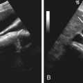
Endoluminal (transvaginal or transrectal) ultrasonography and magnetic resonance imaging (MRI) permit comprehensive evaluation of urethral diverticula, including assessment of size, number, location, and configuration ( Figure 79-3 ).

Ultrasonography is performed using a broadband 5- to 10-MHz linear array for transperineal scanning (transducer placed on the labia) and a broadband 5- to 9-MHz curved array for transvaginal scanning (transducer placed 1 to 2 cm in the vaginal introitus).
MRI for assessment of urethral diverticula may be performed with a surface or endoluminal (endovaginal or endorectal) coil. Transvaginal MRI is performed using a disposable endovaginal coil after intramuscular injection of 1 mg of glucagon (to prevent rectal contractions), axial and sagittal fast spin echo T2-weighted imaging, and unenhanced and gadolinium-enhanced axial spin echo T1-weighted imaging.
Urethral diverticula occur most frequently along the posterolateral wall of the mid-urethra. They appear as cystic paraurethral lesions on imaging ( Figures 79-4 through 79-6 ). The diverticular neck is usually best demonstrated on endoluminal MRI. Fluid/fluid levels, internal echoes, or altered signal intensity may result from hemorrhage or superimposed infection. Malignant degeneration manifests as soft tissue mass within the diverticulum in this situation.


Treatment
Small asymptomatic diverticula may be managed expectantly. Acutely inflamed diverticula should be initially treated with antibiotics. Symptomatic or large urethral diverticula should be resected. Endoluminal MRI or ultrasonography provides a useful roadmap by accurately identifying the diverticular configuration and neck. Recurrence may occur in up to 30% of patients, usually owing to inadequate excision of the diverticular neck.
Urethritis
Acute urethritis in women is most frequently gonorrheal (Neisseria gonorrhoeae) or trichomonal (Trichomonas vaginalis) and less commonly is due to infection by Chlamydia trachomatis. It is characterized clinically by dysuria, frequency, and nocturia. Diagnosis is usually based on clinical and laboratory workup. The role of imaging is limited in the acute phase ( Figure 79-7 ). Imaging plays a role in diagnosing complications such as postinflammatory urethral strictures and evaluation of the upper urinary tract for involvement by infection.


