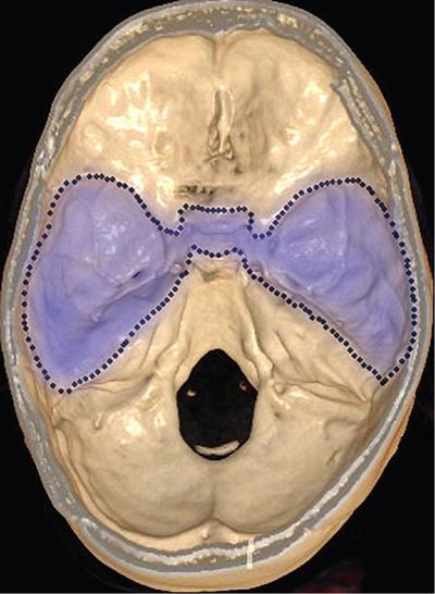
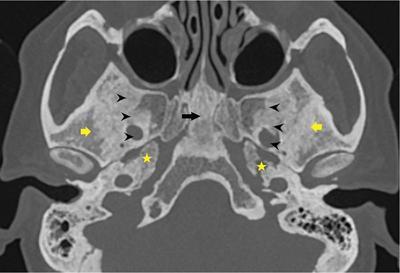
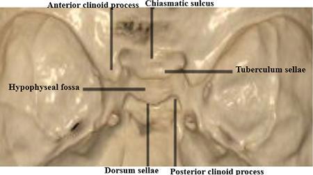
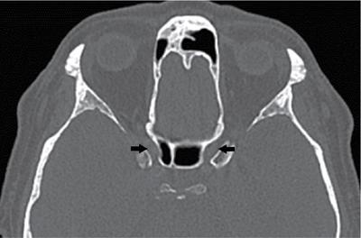
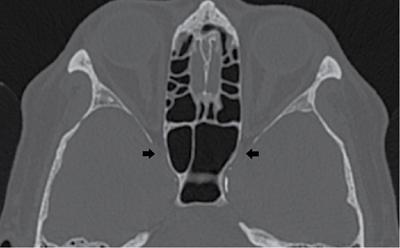
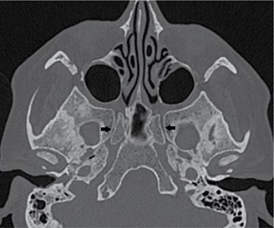
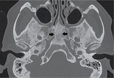
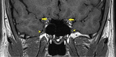
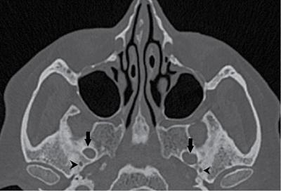
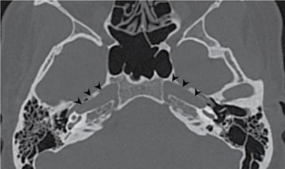
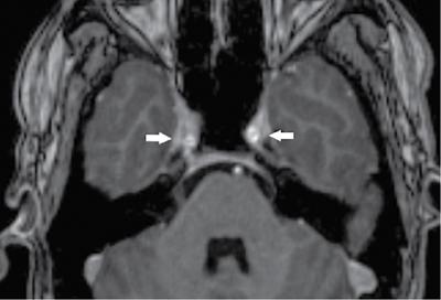
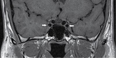
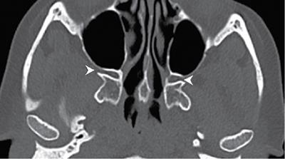
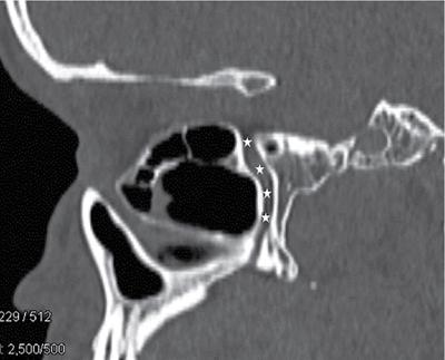
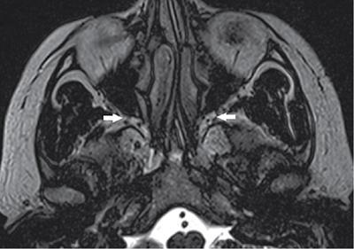
S. Rajakokila, Aarathhi Dhevi Vikramvel, Subarekha TB The middle cranial fossa is located centrally in the floor of cranial cavity. It has a “butterfly-shaped configuration” with middle segment accommodating the pituitary gland and two lateral segments accommodating temporal lobes of the brain. It consists of three bones – the sphenoid bone and two temporal bones (Fig. 3.27.1). Anteriorly, it is bounded by the limbus of sphenoid bone. The limbus is a bony ridge which forms the anterior margin of chiasmatic sulcus (a groove running between right and left optic canals). Anterolaterally, it is bounded by lesser wings of the sphenoid bone. Posteriorly, it is bounded by dorsum sellae of the sphenoid bone. Posterolaterally, it is bounded by the superior border of petrous portion of temporal bone. The floor of middle cranial fossa is formed by central skull base, which consists of the body and greater wing of sphenoid, and the squamous and petrous portions of temporal bone. The central segment of middle cranial fossa consists of the pituitary gland, and two lateral segments of middle cranial fossa accommodate the temporal lobes of brain. Numerous bony landmarks are present in the central skull base, as discussed in the following (Fig. 3.27.2). The body of sphenoid bone constitutes the central segment of the middle cranial fossa. It consists of the sella turcica (Latin for Turkish saddle), which is a saddle-shaped bony prominence. It holds and support the pituitary gland and consists of three parts: The sella turcica is bounded by anterior and posterior clinoid processes. The anterior clinoid processes arise from lesser wing of sphenoid bone. The posterior clinoid processes are superolateral projections of dorsum sellae and provide attachment to tentorium cerebelli (Fig. 3.27.3). The depressed lateral portions of middle cranial fossa are formed by greater wings of sphenoid bone, and the squamous and petrous portions of temporal bone. They support the temporal lobes of the brain. It is the site of many foramina which transmit nerves and vessels. There are many foramina that transmit vessels and nerves in and out of the middle cranial fossa. Chiasmatic groove is a linear depression in transverse plane in the planum sphenoidale and is formed by optic chiasm. Anterior and lateral to optic chiasm lies the optic canal in the medial aspect of lesser wing of sphenoid. Optic nerve (CN II) and ophthalmic artery pass through the optic canal (Fig. 3.27.4). The superior orbital fissure lies inferolateral to optic canal and opens anteriorly into the orbit (Fig. 3.27.5). It is situated between lesser and greater wing of sphenoid. It allows oculomotor nerve (CN III), trochlear nerve (CN IV), abducens nerve (CN VI), ophthalmic division of trigeminal nerve (CN V1) and superior ophthalmic vein and sympathetic fibres. The foramen rotundum lies more inferiorly in a canal-like orientation and opens into the pterygopalatine fossa (Fig. 3.27.6). The maxillary division of trigeminal nerve (CN V2) traverses the foramen rotundum. It also provides communication between the cavernous sinus and pterygopalatine fossa. The inferior orbital nerve, terminal branch of V2 extends from pterygopalatine fossa to inferior orbital fissure. The vidian canal lies in the greater wing of sphenoid, more inferiorly and medially to foramen rotundum, and consists of vidian nerve/artery (Fig. 3.27.7). The posterior opening of vidian canal lies at foramen lacerum and anteriorly extends to the pterygopalatine fossa (Fig. 3.27.8). The foramen ovale lies more laterally within the greater wing of sphenoid and opens into the infratemporal fossa. It also acts as a conduit to masticator space. It gives passage to the mandibular division of trigeminal nerve (CN V3), lesser petrosal nerve and accessory meningeal branch of internal maxillary artery (Fig. 3.27.9). The foramen spinosum is located posterolateral to foramen ovale in the greater wing of sphenoid and also opens into the infratemporal fossa. It transmits middle meningeal artery, vein (Fig. 3.27.9). The carotid canal is located posteriorly and medially to the foramen ovale. This is traversed by the internal carotid artery which enters the cranium to the supply the brain parenchyma (Fig. 3.27.10). At the junction of sphenoid, temporal and occipital bones lies the foramen lacerum. It is filled with cartilage and acts as a floor to lacerum segment of ICA (Fig. 3.27.12). Neurovascular foramen at the central skull base and structures traversing these foramina are summarized in Table 3.27.1. Cavernous sinus and pterygopalatine fossa are two main sites providing easy communication between intra- and extracranial compartments. These are paired dural-based sinuses lying on either side of body of sphenoid bone. They receive venous drainage from ophthalmic veins and sphenoparietal sinuses and drain into superior and inferior petrosal sinuses (Fig. 3.27.11). The lateral margin of cavernous sinus is concave. The lateral wall consists of cranial nerves CN III, CN IV, CN V1 and CN V2. ICA and CN VI lie medially. Extracranial communications: It has an inverted pyramid configuration. It extends between inferior orbital fissure superiorly and palatine foramen inferiorly, which opens into posterior aspect of oral cavity (Figs. 3.27.13 and 3.27.14). It consists of sphenopalatine ganglion, branches of CN V2 and distal internal maxillary artery (Fig. 3.27.15). To aid in narrowing the differentials, the central skull base can be divided into three compartments:
3.27: Imaging of middle cranial fossa and fifth cranial nerve (trigeminal nerve)
Abbreviations
Part A: Middle cranial fossa
Imaging anatomy
Boundaries
Contents
Central segment
Lateral segment
Neurovascular foramen
Foramen
Contents
Optic canal
CN II, ophthalmic artery
Superior orbital fissure
CN III, CN IV, CN V1, CN VI, superior ophthalmic vein
Inferior orbital fissure
Inferior orbital nerve, artery and vein
F. rotundum
CN V2, artery of foramen rotundum, emissary veins from cavernous sinus to pterygoid plexus
F. ovale
CN V3, accessory meningeal branch of internal maxillary artery, lesser petrosal nerve
F. spinosum
Middle meningeal artery
Vidian canal
Vidian nerve and artery
Carotid canal
ICA and sympathetic plexus
F. lacerum
Pseudoforamen, cartilaginous floor of lacerum segment of ICA
Important landmarks
Cavernous sinus
Contents.
Pterygopalatine fossa
Contents.
Main connections
Pathology
Central skull base pathology
Midline sagittal central skull base
Related posts:
![]()
Stay updated, free articles. Join our Telegram channel

Full access? Get Clinical Tree


Radiology Key
Fastest Radiology Insight Engine


