Technical Aspects
Scrotal imaging has been one of the undeniable success stories of modern radiology. The scrotum is predominantly imaged for two clinical indications: the painless scrotal mass and the acute scrotum. Both conditions predominantly affect young men in the second through fourth decades of life. Rapid and accurate diagnosis is the goal of all imaging. Among several imaging modalities available, ultrasonography and magnetic resonance imaging (MRI) are used predominantly, whereas computed tomography (CT), angiography, and nuclear medicine studies are rarely used as primary imaging modalities for disorders of the scrotum.
Ultrasonography
Ultrasonography is undoubtedly the mainstay and is invariably the first and often the only imaging modality necessary. However, MRI can be useful as a problem-solving tool when ultrasonographic findings are equivocal or suboptimal.
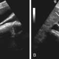
Scrotal ultrasonography is performed with the patient in the supine position and the scrotum supported by a rolled towel placed between the thighs. Optimal results are obtained with a high-frequency (7- to 10-MHz) linear-array transducer. Scanning is performed most often with the transducer in direct contact with the skin, but, if necessary, a stand-off pad can be used for evaluation of superficial lesions.
The testes are examined in at least two planes, along transverse and long axes. The size and echogenicity of each testis and epididymis are compared with those of the opposite side. Color Doppler and pulsed-wave Doppler imaging parameters are optimized to display low-flow velocities to demonstrate blood flow in the testes and surrounding scrotal structures. Power Doppler imaging also may be used to demonstrate intratesticular flow in patients with an acute scrotum. In patients being evaluated for an acute scrotum, the asymptomatic side should be scanned initially to set the gray-scale and color Doppler gain settings to allow comparison with the affected side. Transverse images with portions of each testis on the same image should be acquired in gray-scale and color Doppler modes. Scrotal skin thickness is evaluated. The structures within the scrotal sac are examined to detect extratesticular masses or other abnormalities. In patients with small palpable nodules, scans should include the area of clinical concern. A finger should be placed beneath the nodule and the transducer placed directly over the nodule for scanning. Alternatively, a finger can be placed on the nodule and the transducer opposite to allow visualization of the lesion. Additional techniques such as use of the Valsalva maneuver or upright positioning can be used as needed for venous evaluation.
Magnetic Resonance Imaging
For MRI of the scrotum, a folded towel is placed between the patient’s legs to elevate the scrotum and penis. A local surface coil is used. The penis is dorsiflexed over the pubis and a pad placed over it to retain its position. It is important to take time with positioning and ensure patient comfort, because any movement either voluntary or from cremasteric contraction may degrade image quality. A typical imaging protocol consists of large field-of-view (FOV) axial pelvic imaging to assess the inguinal canal and bowel for hernias and to detect ascites. The origins of the renal vessels should be included to allow evaluation of the lymph node drainage of the testes. A high-resolution T2-weighted fast spin-echo sequence (FOV of 10 to 12 cm, imaging matrix 256 × 256) is used in the axial, sagittal, and coronal planes to image the scrotum. A high-resolution, axial, T1-weighted spoiled gradient echo sequence may be used to identify hemorrhage.
Gadolinium-enhanced imaging is not routinely used but can be performed in selected instances. Use of contrast material can aid in differentiating between a benign cystic lesion and a cystic neoplasm and can help assess for areas of absent or reduced testicular perfusion, such as in segmental testicular infarct. When gadolinium-enhanced imaging is indicated, a fat-saturated T1-weighted spoiled gradient echo sequence is obtained.
Pros and Cons
Modern ultrasonography provides high-resolution images of the scrotum and its contents, is relatively inexpensive and quick to perform, has no known bioeffects, and allows dynamic maneuvers such as the Valsalva maneuver to be incorporated into the examination. However, ultrasound imaging requires skill and is therefore operator dependent.
MRI provides excellent tissue detail. Contrast-enhanced MRI may allow diagnosis of some benign masses that are indeterminate on ultrasonography. MRI also may aid detection of undescended testes that are not visualized on ultrasonography. In the acute scrotum, MRI can help diagnose testicular torsion; and when dynamic contrast-enhanced MRI is used in combination with T2- and T2*-weighted images, testicular necrosis may be diagnosed. MRI is more expensive, takes slightly longer to perform, and cannot be used in patients with pacemakers, implants, and claustrophobia.
Controversies
The field strength of MRI used in clinical practice and diagnostic radiofrequency coils are regarded as safe, and no harmful effects have been recorded. However, the potential effects are of some concern, because radiofrequency radiation increases tissue temperature and spermatogenesis is particularly susceptible to temperature-related impairment. Conflicting reports exist about the effects of MRI on the testis itself. However, it is unlikely that the relatively mild transient increase in scrotal temperature produced by a routine diagnostic MRI examination will cause significant damage. Thus, MRI should be used when there is a significant diagnostic advantage over ultrasonography.
Normal Anatomy
The normal adult testis is ovoid and measures 3 cm in anteroposterior dimension, 2 to 4 cm in width, and 3 to 5 cm in length. Each testis normally weighs between 12.5 and 19.0 g. Both the size and weight of the testes normally decrease with age.
On ultrasonography the normal testis is slightly echogenic with a homogeneous echotexture. The testis is surrounded by a fibrous band, the tunica albuginea, which is often not visualized in the absence of intrascrotal fluid ( Figure 77-1 ). The tunica is often seen as an echogenic structure where it invaginates posteriorly into the testis to form the mediastinum.

The epididymis is a tubular curved structure consisting of a head, body, and tail that lies posterolateral to the testis and measures 6 to 7 cm in length. On ultrasonography, it is isoechoic to hyperechoic to the normal testis and has equal or diminished vascularity. The epididymal head is the largest and most easily identified portion of the epididymis. It lies superolateral to the upper pole of the testis and is an important landmark during the ultrasound examination ( Figure 77-2 ). It is composed of 8 to 12 efferent ducts converging into a single larger duct in the body and tail. This single duct becomes the vas deferens and continues in the spermatic cord ( Figure 77-3 ).


