Imaging Soft Tissues of the Neck


Imaging of soft tissues of the neck can be essential in the evaluation of patients with a variety of chief complaints, including neck trauma, ingested or aspirated foreign body, nontraumatic neck pain and swelling, dysphagia and voice change, visible or palpable mass, and central nervous system complaints with possible vascular causes. The neck contains vascular, nerve, airway, gastrointestinal, and bony structures, any of which may be the source of pain. Remember that referred pain from other regions of the body may present with neck pain, so a broad differential diagnosis should be entertained in formulating an imaging plan. Fascial planes connect compartments of the neck with the mediastinum and thoracic prevertebral spaces, posing a risk of spread of infection from the neck to these regions. Imaging of the neck often is performed with imaging of the head or chest, as structures passing through the neck extend into these adjacent body regions. In this chapter, we explore the modalities available for soft-tissue cervical imaging, discuss clinical indications for imaging in a variety of chief complaints, and review some characteristic findings of important pathology, using the figures throughout the chapter. Imaging of the soft tissues of the cervical spinal cord and ligaments are discussed in Chapter 3 .


Who Needs Soft-Tissue Imaging? Which Imaging Modality Should Be Used?


Given the range of potential pathology discussed earlier, it should come as little surprise that no single clinical decision rule can be used to inform decisions for soft-tissue neck imaging. Instead, the differential diagnosis under consideration should drive the imaging decision, based on expected features of the pathology and the capabilities of each modality. Dilemmas for the practitioner include whether imaging is required and, if so, whether to screen with a limited test such as x-ray, rather than incurring the additional cost and radiation exposure of computed tomography (CT). Some soft-tissue neck abnormalities are best assessed with neither x-ray nor CT but rather with ultrasound, magnetic resonance imaging (MRI), fluoroscopy, or techniques such as bronchoscopy and esophagoscopy. First we consider some general principles regarding the available imaging modalities: x-ray, CT, ultrasound, and MRI. Fluoroscopy is discussed later with regard to esophageal pathology.


Soft-Tissue X-ray of the Neck


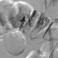
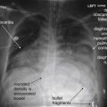
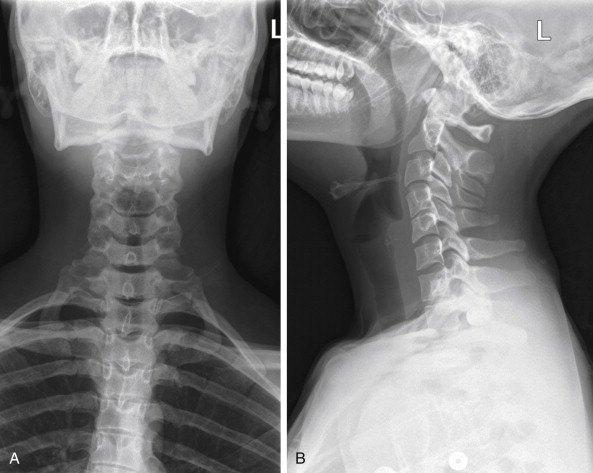
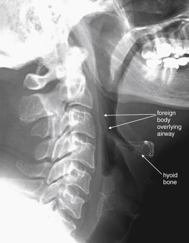
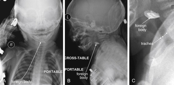
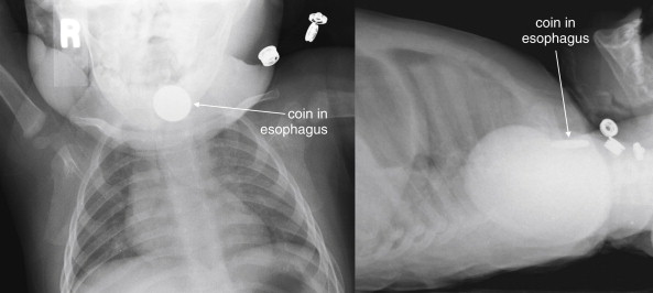
Plain x-ray ( Figures 4-1 and 4-2 ) provides limited information about the soft tissues of the neck. X-ray relies on differentiation of adjacent structures using four basic tissue densities: air, fat, water (which includes soft tissues, both solid organs such as muscle and fluids such as blood), and bone (sometimes called metal density ). As we discuss in detail in Chapter 5 with regard to chest x-ray, two adjacent structures with the same basic tissue density are indistinguishable on x-ray; no border is seen separating them. When two tissues of different density abut one another, the transition is clear. For this reason, x-rays can demonstrate abnormal soft-tissue air ( Figures 4-3 to 4-5 ), deviation or compression of normal air-filled structures (the trachea particularly) ( Figures 4-3 and 4-6 to 4-10 ), air–fluid levels suggesting abscess, and radiopaque foreign bodies ( Figures 4-11 to 4-15 ), as all of these involve a contrast between two key tissue densities. Unfortunately, many of the soft-tissue abnormalities of interest to us as emergency physicians may not involve such a contrast. Instead, one soft-tissue structure may abut a second soft-tissue structure, and these are indistinguishable on x-ray. This leaves an array of potentially devastating forms of neck pathology that are poorly assessed with x-ray. Examples include vascular dissections, abscesses, and subtle soft-tissue masses that may not be seen on x-ray.


Figure 4-1


Normal soft-tissue neck x-ray.

A soft-tissue neck series consists of an anterior–posterior (AP) (A) and a lateral (B) x-ray of the neck. Compared with a cervical spine x-ray, the images are intentionally underexposed to allow soft tissues to be examined. Figure 4-2 examines close-ups from these images.


Figure 4-2


Normal soft-tissue neck x-ray.

A, Anterior–posterior view. B, Lateral view. C, Close-up focusing on epiglottis. Compare with later figures showing pathology. This young adult has a normal x-ray. Note the normal epiglottis and vallecula, the position of the hyoid, and the thickness of the normal retropharyngeal (prevertebral) spaces. These become abnormal in disease states such as croup, epiglottitis, and retropharyngeal abscess.


Figure 4-3


Retropharyngeal abscess with retropharyngeal air.

This 22-month-old boy presented with drooling and hyperextension of the neck. He had been seen days earlier and diagnosed with a likely viral pharyngitis. A, Lateral soft-tissue x-ray of the neck shows extensive air (black) dissecting into tissue planes of the deep spaces of the neck, as well as the submental area. The retropharyngeal space is dramatically wide with visible air and is deviating the airway forward. Classically, soft-tissue x-ray in the setting of retropharyngeal abscess may also reveal an air–fluid level in the retropharyngeal space—a finding not present in this image. B, Normal soft-tissue x-ray is shown for comparison. The next figure demonstrates the computed tomography images from this patient. The patient was intubated and ultimately taken to the operating room for surgical drainage, which confirmed abscess.


Figure 4-4


Retropharyngeal abscess: CT with intravenous (IV) contrast, soft-tissue windows.

Same patient as Figure 4-3 . Dramatic dissection of gas (black) is seen in the retropharyngeal space. A, The endotracheal tube is visible. Air outlines the carotid and internal jugular vessels. B, Air is seen dissecting into the superior mediastinum. C, Gas is seen medial to the aortic arch. This case highlights the dangers of retropharyngeal processes, as infections in this location can readily extend to the mediastinum. Interestingly, the patient’s infection was caused by yeast, and the extensive gas collection may have been carbon dioxide produced by the Candida species.


Figure 4-5


Retropharyngeal air.

This 10-year-old boy complained of chest pain and dysphagia after coughing. A, Lateral soft-tissue neck x-ray reveals a thin stripe of air in the retropharyngeal space, extending from the skull base to the chest. Given the clinical history, the airway or lungs were the likely source of the air leak. Compare with the normal soft-tissue x-ray in a Figure 4-1 , B. The patient underwent computed tomography (CT) of the neck and chest with intravenous contrast but the source of the air was not explicitly identified. B, Neck CT (shown on soft-tissue windows) shows a thin stripe of retropharyngeal air, correlating with the soft-tissue x-ray. Noncontrast CT could have revealed this finding, although contrast was given because of the possibility of an infectious source that might enhance with contrast.


Figure 4-6


Retropharyngeal hematoma with airway compromise.

This 67-year-old woman presented with neck pain after tripping, falling forward, and striking her nose against a windowsill. Although she did not lose consciousness, she reported a popping sound from her neck as hyperextension occurred. During computed tomography (CT) scan of the head and cervical spine, the patient developed stridor and was immediately removed from the CT scanner. Within 1 minute, she experienced a respiratory arrest and was intubated with difficulty. CT scans before and after her intubation are shown in the next figure. In this figure, lateral computed tomography (CT) scout images are shown, as these approximate the findings from a lateral x-ray, had one been performed. A, Scout CT image performed before the patient complained of dyspnea shows an extremely wide retropharyngeal space and airway compression. B, Normal scout film is shown for comparison.

Beware of placing a patient supine for CT, because this can worsen airway compromise.


Figure 4-7


Retropharyngeal hematoma with airway compromise.

Same patient as Figure 4-6 . CT scans before and after her intubation are shown.

A, The caudadmost slice of her noncontrast facial CT shows a dramatic retropharyngeal hematoma displacing the airway forward (double arrow). The normal retropharyngeal space is less than 6 to 8 mm at C1-3 and 18 mm at C6 or C7, while this patient’s hematoma was more than 40 mm in thickness. B, After intubation, the patient underwent repeat CT with intravenous contrast to assess for active hemorrhage—none was found. The retropharyngeal hematoma is still visible, 20 mm in thickness at this level posterior to the endotracheal tube (double arrow). But for the presence of the endotracheal tube, the hematoma completely obscures the airway. The hematoma extended caudad into the mediastinum. The patient had no spinal injury detected, and coagulation studies on the patient were normal.


Figure 4-8


Croup.

This 19-month-old, fully immunized boy presented with acute onset of stridor without fever. The patient’s mother was uncertain whether he might have ingested a foreign body, and x-rays of the soft tissues of the neck and chest were obtained. A, The anterior–posterior neck x-ray shows a classic example of the long-segment subglottic narrowing typical of croup, called the “steeple sign” for its resemblance to a traditional peaked church steeple. B, The prevertebral soft tissues are normal, and the epiglottis appears normal on lateral x-ray. The hypopharynx appears ballooned, which is common in croup. X-rays are not needed routinely to confirm croup when a typical history and physical is present. The primary purpose of x-rays in this setting is to rule out other etiologies, such as foreign body aspiration or epiglottitis.


Figure 4-9


Epiglottitis.

A, Lateral cervical soft-tissue x-ray showing epiglottitis in a 64-year-old man. The epiglottis is thickened, showing a classic “thumb sign.” B, Normal soft-tissue x-ray for comparison. The brightness and contrast have been increased to point out the normal thin epiglottis, which is partially hidden behind the hyoid bone. The vallecula is seen as a narrow space anterior to the epiglottis. An anterior–posterior (AP) x-ray is not shown for this case, because the AP view is not usually diagnostic in cases of epiglottitis.


Figure 4-10


Epiglottitis, seen on CT with IV contrast, soft tissue windows.

While a patient with suspected impending airway occlusion should not be placed supine in a CT scanner, CT can be a valuable tool for diagnosis of airway and retropharyngeal pathology. In this patient with epiglottitis, the CT scout image (A) demonstrates a thumb sign similar to the plain film findings in Figure 4-9 . The axial image (B) also demonstrates an enlarged epiglottis, with an obscured left piriform sinus. A normal epiglottis in another patient is shown for comparison (C). The normal epiglottis is a thin crescent.


Figure 4-11


Penetrating neck trauma.

This 30-year-old man was wounded with a shotgun. The patient ducked his head at the moment of the blast, and most of the shot impacted superficially in his scalp. However, one pellet entered the mental vertex of his mandible and lodged in the anterolateral neck. A, Anterior–posterior view shows the pellet to be in the lateral left neck, in the region of the carotid artery. B, The lateral x-ray combined with the AP view shows the pellet is not superficial. Computed tomography was performed to assess the carotid artery and adjacent structures Figure 4-12 .


Figure 4-12


Penetrating neck trauma.

Same patient as Figure 4-11 . Computed tomography with intravenous contrast was performed to assess the carotid artery and adjacent structures. A, Soft-tissue windows. B, Bone windows. The pellet creates extensive metallic streak artifact, which completely obscures the vessels of interest. Conventional angiography could be performed in circumstances such as these, because it is free of this type of artifact.


Figure 4-13


Airway foreign body: Chicken bone.

This 47-year-old man was unable to speak after eating chicken. The patient pointed to his larynx, indicating a foreign body sensation. Lateral neck x-ray shows a foreign body superior to the larynx, perpendicular to the hyoid bone. Remember that you must look at both the anterior–posterior (AP) and the lateral view to localize an object; in this case, however, the object was not visible on the AP view, because it overlay the spine.

Smaller foreign bodies such as fish bones may not be visible on plain film but may be identified on computed tomography. In this case, the patient was taken to the operating room for removal of the object, which was found to be a 4-cm chicken bone in the right piriform sinus.


Figure 4-14


Airway foreign body: Metal mesh.

The role of plain films in evaluation of soft tissues of the neck is limited, but when the patient’s airway is threatened, plain film may offer the only safe imaging modality. Portable plain films with the patient in a “position of comfort” may offer some diagnostic information. CT in a supine position may further compromise airway patency.

In this toddler, parents reported sudden stridor but did not witness an ingestion or aspiration. Portable x-ray revealed a radiopaque mesh object in the airway. The patient was taken emergently to the operating room for removal. Plastic or organic material may not be visible on plain x-ray. A, Anterior–posterior view. B, Lateral view. C, Close-up from B.


Figure 4-15


Esophageal foreign bodies.

This 11-month-old presented with drooling after being witnessed to place a penny in his mouth. X-ray confirms a circular object apparently lodged in the esophagus. A, Anterior–posterior (AP). B, Lateral. Coins lodged in the esophagus tend to assume an “en face” position, with a circular appearance on the AP view (think British oesophagus for the O appearance of an esophageal coin). Coins lodged in the trachea tend to assume an “edge-on” linear appearance on the AP view, apparently directed to that position by the incomplete tracheal rings. This patient was taken to the operating suite for removal of the coin, which was found to be lodged in the esophagus at the level of the cricopharyngeus.


Figure 4-16


Peritonsillar abscess.

A classic peritonsillar abscess requires no imaging, as it has a characteristic appearance on physical examination, with a deviated uvula and peritonsillar mass. In reality, some patients present with early abscesses that are not visible, and others experience too much trismus to allow a complete exam. If more information is needed and the airway appears sufficiently patent for the patient to lie supine safely, intravenous (IV) contrast-enhanced computed tomography can provide excellent diagnostic information. IV contrast leads to “ring enhancement,” an increase in the density on the periphery of an abscess caused by increased blood flow.

A, A small peritonsillar abscess is beginning to form, visible as a hypodense area with slight surrounding enhancement. B, A larger hypodense abscess has formed and is deviating the airway.


Figure 4-17


Anterior neck abscess, possibly arising from a dental infection.

In this soft-tissue neck computed tomography with intravenous contrast viewed on soft tissue windows, a hypodense abscess containing fluid ( dark gray ) and air ( black ) is present in the patient’s right neck in a submandibular position. A, Soft-tissue changes including swelling and inflammatory stranding ( smoky appearance ) of subcutaneous fat are present (compare with opposite side). B, A tiny fleck of air is visible just medial to the mandible, suggesting that infection may have arisen from a mandibular tooth.


What Information Can Be Gleaned from a Portable Soft-Tissue Neck X-ray? How Should It Be Interpreted?


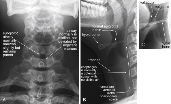
Portable anterior–posterior (AP) and lateral soft-tissue neck x-rays provide basic information about the airway, as described earlier. Like a focused assessment with sonography for trauma (FAST), the soft-tissue neck film should be assessed by the emergency physician with several specific clinically relevant questions in mind. As you interpret an x-ray, ask yourself each of these questions, rather than simply looking at the x-ray, and assess the x-ray for relevant findings.


On the AP x-ray:


  • 1.

    Is the airway deviated laterally or narrowed, suggesting an extrinsic mass?


  • 2.

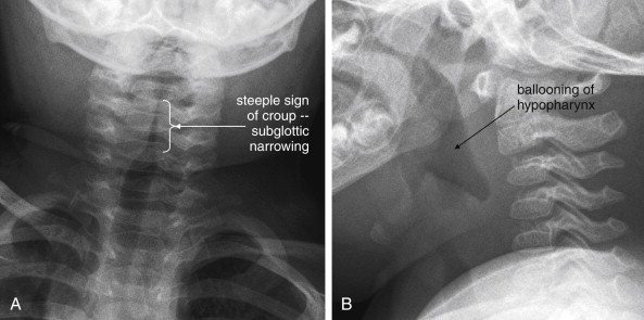
    Is the airway narrowed by concentric inflammation, producing a “steeple sign” characteristic of croup (see Figure 4-8 )?


  • 3.

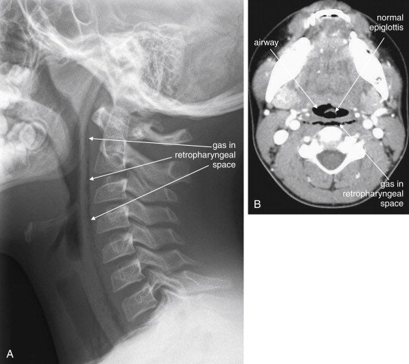
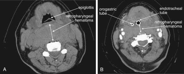
    Is air visible in the soft tissues of the neck, indicating potential pneumomediastinum, prevertebral air, esophageal or tracheal perforation, or retropharyngeal abscess?


  • 4.

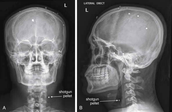
    Is a radiopaque foreign body visible (see Figures 4-11 and 4-14 )?


On the lateral x-ray:


  • 1.

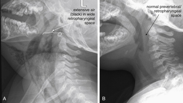
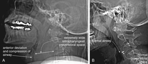
    Is the airway deviated or narrowed in the AP direction? If so, why? Is an anterior neck mass compressing the trachea, or is a retropharyngeal process exerting a mass effect ( see Figure 4-6 )?


  • 2.

    Are the soft tissues directly behind the trachea in the retropharyngeal and prevertebral spaces thickened? If so, do they impinge upon the airway (see Figures 4-3, 4-5, and 4-6 )? The normal thickness of prevertebral spaces has been reported in several studies, using both x-ray and CT ( Table 4-1 ).


    TABLE 4-1

    Normal Prevertebral Soft-Tissue Thickness


    Imaging Modality and Location Thickness
    X-ray
    Retrocricoid soft-tissue thickness (x-ray) 0.7× diameter of C5 vertebral body
    Retrotracheal soft-tissue thickness (x-ray) 1.0× diameter of C5 vertebral body
    CT
    C1 8.5 mm
    C2 6 mm
    C3 7 mm
    C4-5 Not determined because of variable position of esophagus and larynx
    C6 18 mm
    C7 18 mm

    From Chen MY, Bohrer SP. Radiographic measurement of prevertebral soft tissue thickness on lateral radiographs of the neck. Skeletal Radiol 1999;28(8):444-6; Rojas CA, Vermess D, Bertozzi JC, et al. Normal thickness and appearance of the prevertebral soft tissues on multidetector CT. AJNR Am J Neuroradiol 30(1):136-41, 2009.


  • 3.

    Is an air–fluid level present posterior to the trachea, representing an abscess that could rupture into the airway or extrinsically compress it?


  • 4.

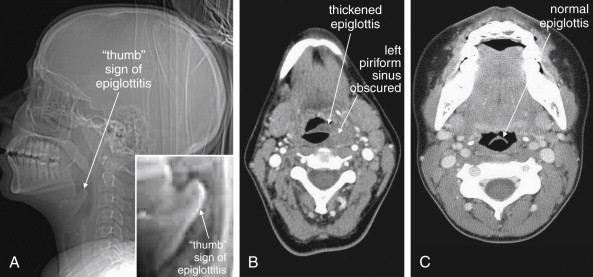
    Is the epiglottis enlarged, threatening supraglottic airway obstruction from epiglottitis ( see Figures 4-9 and 4-10 )?


  • 5.

    Is abnormal air visible in the neck, indicating potential pneumomediastinum, prevertebral air, esophageal or tracheal perforation, or retropharyngeal abscess (see Figures 4-3 and 4-5 )?


  • 6.

    Is a radiopaque foreign body visible (see Figures 4-11 and 4-13 to 4-15 )?


Soft-Tissue Neck CT


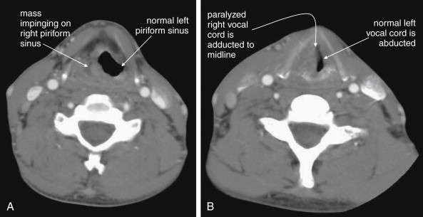
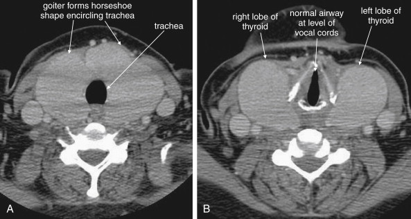
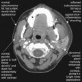
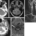
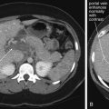
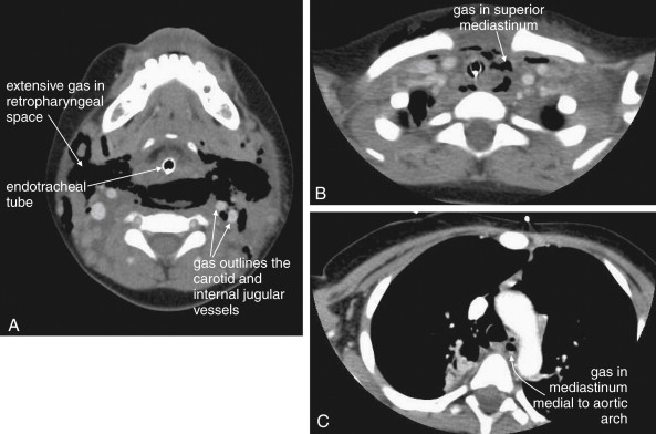
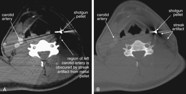
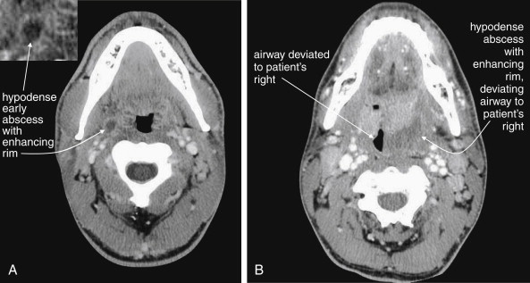
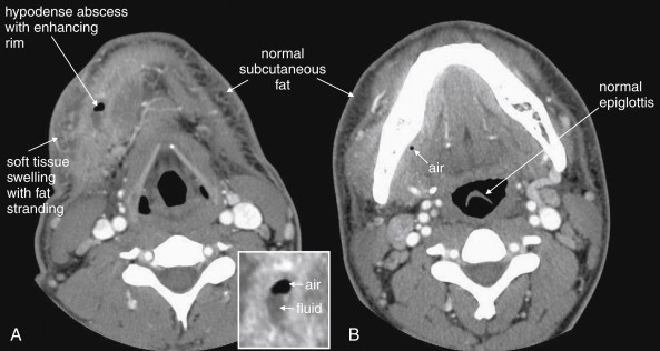
CT is clearly superior to x-ray, as it is able to distinguish a subtler spectrum of tissue densities. CT scan readily distinguishes air, fat, fluid, solid soft tissues of muscle density, and bone. The window setting on CT (discussed in detail later) can be adjusted to highlight a particular tissue density, but on all window settings these tissues are color coded from black (lowest density) to white (highest density). On soft-tissue windows, which are key in our evaluation of neck soft-tissue pathology, air is black, fat is a dark gray (nearly black), fluids such as blood or pus are an intermediate gray, solid tissues are a slightly lighter gray, and high-density material such as bone or metal are white. Lung window settings are sometimes used to evaluate the neck, as air becomes quite evident (black) against all other tissue densities, which appear white on this window setting. Bone windows also highlight air (black) in contrast to all other tissues. Nonstandard window settings can be selected to highlight pathology. The precise density of a tissue can also be measured on CT scan on most picture archiving and communication systems (PACSs) (the name given to a digital diagnostic imaging interface). The computer cursor can be placed over a single pixel or a broader region of interest, and the density of the tissue in Hounsfield units (HU) is reported. On the Hounsfield scale (Figure 000), water has a density of 0 HU, air has a density of −1000 HU, and the highest-density bone has a density of +1000 HU. Nonbiological high-density substances such as metal foreign bodies have densities exceeding +1000 HU. Fat is less dense than water and has a lower density, around −50 HU. Soft tissues such as muscle are denser than water and have a higher Hounsfield value, around 30–50 HU. Liquid blood has a density slightly greater than water, due to its cellular content; the density may vary with factors such as hematocrit and volume status. CT allows ready identification of a tissue abnormality such as abscess based on density differences. An abscess ( Figures 4-16 and 4-17 ) filled with liquid pus appears darker than denser surrounding soft tissues viewed on soft-tissue windows.


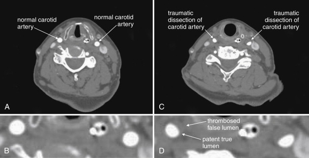
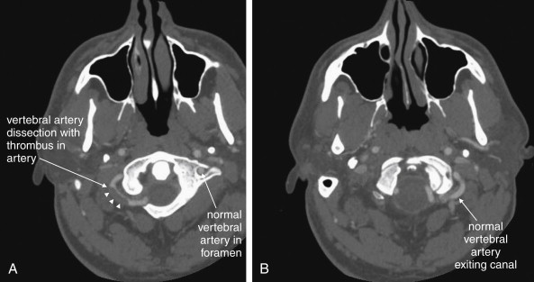
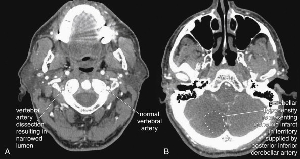
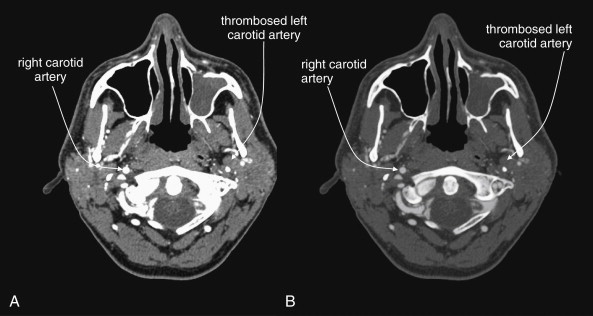
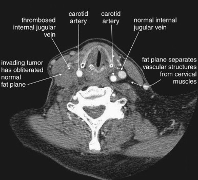
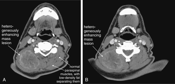
Structures with identical (or nearly identical) density are indistinguishable on CT without contrast, with several clinically important examples. Because pus is similar in density to surrounding normal soft tissues, IV contrast can assist in identifying an abscess. Normal soft tissues are highly vascular and enhance (increase in density) following administration of IV contrast. Pus within an abscess does not enhance with IV contrast, so the difference in density between abscess and normal surrounding tissues increases following IV contrast administration, increasing the conspicuity of the abscess. Similarly a vascular dissection ( Figures 4-18 to 4-20 ) would be difficult or impossible to recognize without contrast, because blood in the true and false lumen share the same density, and the thin intimal flap separating them is of nearly identical density. Administration of intravenous (IV) contrast changes this relationship by providing high-density (contrast) material flowing on both sides of the intimal flap. The low-density flap becomes visible against this background. Partially thrombosed vascular segments ( Figures 4-21 to 4-22 ), which are common in dissections of the relatively small-caliber vessels of the neck, are also made visible by this same principle: high-density contrast flows through the patent lumen of the vessel, making low-density thrombus visible. Without contrast, liquid blood and thrombus share nearly identical densities and are nearly indistinguishable. Contrast also aids in detecting soft-tissue masses. Normal muscle and a muscle tumor such as sarcoma ( Figures 4-23 to 4-25 ) may share the same tissue density without contrast. However, a highly vascular neoplasm receives differentially more parenterally administered contrast and has a higher density when imaged with contrast. CT does not perfectly distinguish benign and malignant mass lesions, so biopsy is necessary for confirmation. Even when an abscess is not present, inflamed or infected soft tissues also have increased blood flow and enhance with IV contrast ( Figure 4-26 ; see also Figures 4-16, 4-17, and 4-26 ). Without any contrast administration, CT is excellent for airway assessment, as very low-density air within the airway provides outstanding intrinsic contrast against surrounding soft tissues. ( Figure 4-27 ; see also Figures 4-7, 4-10, 4-16, 4-17, 4-24, and 4-25 ). However, extreme caution should be applied in sending patients with suspected airway pathology to CT, as airway obstruction can be worsened by supine positioning for CT.


Figure 4-18


Vascular dissections (carotid artery).

This 75-year-old woman presented after a motor vehicle collision. The patient was noted to have an ecchymotic seat belt mark across her left neck. The patient had multiple other injuries, including a diaphragm injury diagnosed by chest x-ray, so CT angiography of the neck was prospectively planned (for assessment of cervical vascular injury) to be performed at the time of chest and abdominal CT to avoid repeating the intravenous contrast bolus. A, The normal carotid arteries below the level of the injury. Note the rounded appearance and bright filling with contrast B, a close-up view from A. C, Bilateral carotid artery dissections at the level of injury. The normal round appearance is replaced by a three-quarter moon appearance. The false lumen has thrombosed bilaterally, so an intimal flap is not seen (compare with aortic dissection in Chapter 7 ). D, a close-up view from C. In some cases, irregularity of the contour of the artery may be the only abnormal finding. The dissection has resulted in significant stenosis, approximately 30% to 40% on the left, and 20% to 30% on the right.


Figure 4-19


Vascular dissections (vertebral artery dissection): CT with IV contrast, viewed on vascular windows.

Spontaneous dissection of the vertebral artery is a relatively rare event but is thought to be one of the most common causes of stroke in young patients. Computed tomography (CT) angiography and magnetic resonance angiography are the most common methods of assessment today, with catheter-based angiography usually reserved for patients with CT or magnetic resonance abnormalities. Remember that the vertebral arteries exit their canals at the C1 level, curve medially, pass through the ring of C1, and then merge to form the basilar artery. In these axial CT images, IV contrast has been administered to the patient. The window level is set to an intermediate value between bone and soft-tissue settings. You can recognize this from the subcutaneous fat, which is not nearly black, and bone, which is too bright to allow its internal structure to be easily recognized. A, The left vertebral artery is seen within its canal. The curving right vertebral artery is seen with a filling defect representing a thrombosed region of dissection. B, The normal left vertebral artery is seen filled with contrast.


Figure 4-20


Vertebral artery dissection from C4 to C1 with posterior inferior cerebellar artery (PICA) territory infarct after cough.

This patient presented with vertigo after paroxysmal cough, and a cervical computed tomography angiogram was performed to evaluate for vertebral artery dissection. A, The left vertebral artery is of normal caliber. The right vertebral artery is narrowed to a fraction of the normal size. Is this a normal variant or acute disease? In congenital hypoplasia of the vertebral artery, the bony canal also is smaller than normal, whereas in this patient the canal is of normal size, indicating that the artery has an acute abnormality. B, A corresponding acute ischemic infarct is seen in the cerebellum in a region supplied by the PICA. Magnetic resonance imaging or magnetic resonance angiography could be used to provide similar information.


Figure 4-21


Carotid artery thrombosis.

This 47-year-old man presented with headache and underwent computed tomography venography of the head and neck to assess for venous sinus thrombosis. Instead, complete occlusion of the left internal carotid artery was discovered. A, Soft-tissue windows. B, A window setting similar to bone windows often used for assessment of vascular structures. The benefit of this window setting is that the internal structure of blood vessels, such as intimal tears, irregularity, or thrombosis is more evident. On typical soft-tissue windows (A), contrast in the vessel is so bright that internal structural detail is often less visible. In this particular case, the pathology is subtle for a different reason—you must recognize the absence of a normal structure in the image. Use expected symmetry to help you. The right carotid artery is well opacified with intravenous contrast. The left carotid artery is devoid of contrast because it has completely thrombosed. On a picture archiving and communication system, follow blood vessels through multiple slices to inspect them. Incidentally, the patient’s left maxillary sinus is opacified, suggesting sinusitis (see Chapter 2 for more information).


Figure 4-22


Internal jugular vein occlusion, CT with IV contrast, soft tissue windows.

This patient with an aggressive esophageal tumor has cervical adenopathy resulting from tumor metastases. The enlarging tumor burden has created local mass effect on the right internal jugular vein, which has become thrombosed as a result. The internal jugular contains dark thrombus on the right, compared with the bright contrast–filled left internal jugular vein. The cervical fat planes on the right have become obscured by invading tumor.


Figure 4-23


Neck mass.

This 70-year-old patient presented with progressive right posterior neck swelling over a period of 1 month following a dental procedure. Asymmetry and a palpable mass were present on exam. Computed tomography (CT) with intravenous contrast was performed to assess for abscess or mass. A heterogeneously enhancing mass is visible in the right posterior paraspinous muscles, measuring approximately 5.2 by 4.2 cm in axial dimension. The mass does not have the appearance of an abscess, which most often would have a lower density (fluid density, darker gray) center. Note how the mass has obliterated the normal planes separating paraspinal muscles—compare this with the patient’s left side, which is normal. Without contrast, these asymmetrical features and the overall size of the mass would be appreciated, but the discrete margins of the mass would not be seen without enhancement because the mass shares the same density with normal muscle. CT does not identify the exact etiology of the mass, although the differential diagnosis for a muscle-density mass lesion includes sarcoma. Biopsy or excision would be needed to prove the diagnosis. Interestingly, the patient underwent fine-needle aspiration that showed gram-negative rods and spindle-shaped cells initially thought to represent reactive myositis or fasciitis—possibly an infection caused by the reported dental procedure. Unfortunately, surgical pathology showed an undifferentiated sarcoma.


Figure 4-24


Neck masses: Laryngeal cancer with vocal cord paralysis, CT with intravenous contrast.

This patient has a laryngeal mass with complete paralysis of the right vocal cord. A, The mass occupies and obscures the left piriform sinus. Without a history of cancer, this appearance could be similar to a soft-tissue infection adjacent to the airway. B, The right vocal cord is completely paralyzed and is therefore is passively adducted to the midline. The left vocal cord is not paralyzed and is thus abducted normally.

Jul 4, 2019 | Posted by in GENERAL RADIOLOGY | Comments Off on Imaging Soft Tissues of the Neck

Full access? Get Clinical Tree

Get Clinical Tree app for offline access