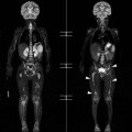
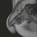
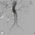
Philip W.P. Bearcroft, Melanie A. Hopper There are numerous imaging investigations available to clinicians and radiologists for the assessment of musculoskeletal disease. This chapter will describe the individual investigations, concentrating on the strengths and weaknesses of each technique. Variations will be discussed and recent imaging advances detailed. A working knowledge of the normal appearance of musculoskeletal structures is essential and these will then be described. Finally, three common imaging scenarios will be explained: calcification in soft tissues, gas in soft tissues and periosteal reaction. Radiography has an important role in the preliminary evaluation of musculoskeletal disorders and for many conditions no further imaging is required. The technique relies upon the differing absorption of ionising radiation by tissues within the body. As a result, radiography provides excellent contrast between tissues such as fat and muscle and bone. It does not, however, provide contrast between soft tissues and has limited ability to demonstrate bone loss compared to cross-sectional techniques. To aid diagnosis, two views of a body part are typically taken. Conventionally these are in the lateral plane and the anteroposterior (AP) plane, which is termed dorsopedal (DP) in the feet and dorsopalmar (DP) in the hands. Particularly in trauma, two views, preferably orthogonal, are important to minimise misinterpretation of overlapping structures (Fig. 45-1). In complex anatomical areas or when specific information is needed, additional or alternative views are frequently obtained. Radiographs are readily available as an imaging technique. Traditionally viewed as hard copy film, technological advances now mean that in the majority of institutions radiographs are obtained, viewed and stored digitally. The picture archiving and communication system (PACS) provides immediate access to previous radiographs for comparison not just for radiography but for all imaging investigations. Radiographs also provide initial soft-tissue assessment and can demonstrate soft-tissue swelling as well as joint effusions, which can be particularly useful in areas where bony abnormality may be radiographically occult (Fig. 45-2). An awareness of the limitations of radiographic evaluation is crucial. As in other areas of the body, artefact from external structures such as clothing and from overlapping structures can be misinterpreted as an abnormality. Some sites can be difficult to appreciate fully using radiographs. The medial end of the clavicle, for example, can be problematic due to multiple overlying structures and an abnormality may be more difficult to detect. It is important that radiographs, as with other imaging investigations, are interpreted in light of the clinical details and additional imaging should be undertaken if concern persists. The lack of radiographic soft-tissue evaluation is well recognised as the attenuation of X-rays by most soft-tissue structures lies within a narrow range and this limits the interrogation of soft-tissue structures. One of the main drawbacks of radiography is its reliance upon ionising radiation and the potential resulting side effects. However, particularly in the extremities, the dose involved is minimal and although the hazards of ionising radiation should never be ignored, the diagnostic benefits generally outweigh the possible risk when radiographs are correctly used. Standard radiographs allow static evaluation of musculoskeletal structures. Imaging a joint under passive or active stress may also provide indirect assessment of ligamentous injury.1,2 For example, comparative flexion and extension views of the cervical spine provide valuable information regarding stability of the atlanto-axial joint (Fig. 45-3). Fluoroscopic techniques have a wide range of uses across radiological specialties. They utilise an X-ray source similar to standard radiographs but provide real-time dynamic video images that are typically used in the radiology department and in orthopaedic surgery to guide interventional procedures such as needle placement and fracture reduction. Images are viewed on a digital unit and are amplified to enable them to be seen in normal light, especially useful in the operating theatre. The basic premise of arthrography is the injection of a radio-opaque contrast medium into a joint usually guided by fluoroscopy, although US guidance can be used as an alternative. Distension of the joint provides indirect information about the soft tissues which can be deduced from the distribution of the injected contrast medium (Fig. 45-4). Fluoroscopic evaluation also allows an element of dynamic assessment. With the advent of more sophisticated cross-sectional imaging which provides detailed and direct assessment of the internal joint structures, arthrography is now more frequently used in combination with MRI and, occasionally, CT following the injection of gadolinium-based or iodine-based contrast medium, respectively. Arthrography is frequently combined with the injection of local anaesthetic to provide additional diagnostic information in patients with possible joint symptoms, helping distinguish assymtpomatic and symptomatic abnormalities; corticosteroid may be introduced as a therapeutic agent. Digital tomosynthesis represents a modification of the conventional radiographic technique to acquire multiple low-dose images of a body part across a range of projection angles of the X-ray tube. Currently well established in breast imaging, interest in the application of tomosynthesis to other areas, including the musculoskeletal system, is growing.3 The radiation dose is greater than that of conventional radiography but less than CT and preliminary studies suggest it has a role in the imaging of orthopaedic prostheses as the metallic-related streak artefacts encountered with CT is reduced.4 Digital tomosynthesis also reduces the difficulty of composite shadow, showing promise in the assessment of anatomically complex areas (Fig. 45-5). In current clinical practice US has an important role in the diagnosis and management of musculoskeletal disease, providing high-resolution assessment of soft tissues. Evaluation of bone is limited, but US can afford information regarding the bony surface and periosteum. Typically, musculoskeletal US requires a linear array high-frequency probe of at least 7 MHz, and greater than 10 MHz is preferred. This is ideal for superficial abnormalities; deeper lesions necessitate a lower-frequency probe to provide adequate depth, albeit at reduced spatial resolution. US main benefits lie in its ability to give real-time, high-resolution and dynamic diagnostic information. In general the technique is fast and well tolerated by patients and allows direct clinical correlation of symptoms and abnormalites by the operator. Importantly, there is no ionising radiation. The ability of US to differentiate between solid and cystic structures is particularly useful in the musculoskeletal system. Soft-tissue calcification is evident earlier on US than on radiographs (Fig. 45-6). Doppler assessment of vascularity is important in soft-tissue masses and also in the assessment of inflammatory and degenerative disorders where the presence of new blood vessel formation, known as neovascularisation, can be used to indicate disease activity and healing (Fig. 45-7). For the assessment of the majority of musculoskeletal conditions, the direction of vascular flow is not particularly useful and so the more sensitive power Doppler (PD) is favoured over colour Doppler techniques, as PD maximises spatial resolution and flow sensitivity at the expense of directional information. The ability of US to demonstrate the movement of tissues in real time underpins one of its main advantages during review of musculoskeletal disorders. Abnormalities such as tendon subluxation and impingement may be clinically suspected but confirmation using real-time US can be invaluable diagnostically and as an aid to treatment. There is widespread use of US guidance for interventional techniques. The anatomy of the extremities in particular allows excellent needle visualisation as it is nearly always possible to position the needle parallel to the transducer, maximising the clarity with which a needle is seen. Ultrasound is operator dependent and there is a steep learning curve. Artefacts can be challenging in musculoskeletal scanning.5 Anisotropy describes the artefactual loss of tissue reflectivity when the US beam is applied at an oblique course to the tissue fibres under examination. The highly structured and fibrillar arrangement of tissues such as tendons and muscle makes this a particular problem in musculoskeletal US (Fig. 45-8A) and the resultant hypoechogenicity can simulate an abnormality such as tendinosis or tearing of a muscle or tendon. Beam edge artefact is evident at the edge of larger tendons such as the Achilles, with loss of normal signal and posterior acoustic shadowing that can mimic or conceal abnormal findings. Beam steering of the transducer array produces tilting of the beam electronically of up to 40° from the main beam direction. When utilised instead of, or in addition to, manual angulation of the probe, this allows image generation from differing angles and provides a reduction in or even elimination of anisotropic artefact (Fig. 45-8). Extended field-of-view (EFOV) or panoramic ultrasound gives a continuous image greater than the size of the ultrasound probe and although this does not necessarily improve the diagnostic ability of US, it provides images which more readily demonstrate the relevant abnormal findings than on the static images obtained by the operator (Fig. 45-9). The US assessment of tissue elasticity as an aid to diagnosis is well established in other radiological sub-specialities such as breast imaging and early studies suggest that there will be a role for the technique in musculoskeletal scanning. Elastography provides an assessment of tissue stiffness and requires the operator to gently compress the tissues under evaluation beneath the transducer. The strain or displacement of the tissues is determined by their hardness or softness. Normal ligaments and tendons are resistant to compression but tissue softening occurs in several conditions including degeneration and trauma.6 The potential of elastography does not lie in the replacement of traditional B-mode US imaging but rather as a complementary technique. There is particular potential in the assessment of soft-tissue conditions which on B- mode imaging have an isoechoic appearance to the normal surrounding tissues such as early tendon degeneration or tearing (Fig. 45-10). Development of abnormal new blood vessels in musculoskeletal pathology is accompanied by the formation of abnormal nerves and there is good scientific correlation between these changes and pain.7 Therefore, the early identification of new vessels is important in the diagnosis of disease and potentially provides a target for treatment (Fig. 45-7). Contrast media-enhanced US (CEUS) detects low blood flow that may not be detectable by more traditional Doppler techniques. The intravenous injection of microbubble contrast media has been shown to allow more accurate assessment of synovial vascularity, which in turn correlates with disease activity in rheumatological disorders.8 How this will influence patient management in the clinical setting is not fully understood and currently the use of CEUS in the assessment and follow-up of rheumatological disease remains at the research level. CT is ideally suited for the evaluation of bony structures and soft-tissue calcification and provides excellent spatial resolution and demarcation of bony structure and detail. Typically, images are acquired in the axial plane but it is a noteworthy strength that CT images can be reconstructed into any other plane. Surface 3D-rendered reformatting can also be a useful aid to surgical planning. Despite its limitations in assessment of muscle and fat, CT has a role in imaging patients in whom MRI is contraindicated and can be combined with the injection of intra-articular iodinated contrast media in a similar way to the more widely utilised MR arthrography. As with radiography, CT relies upon ionising radiation. CT, even of an extremity, is associated with a radiation dose many times that of conventional radiography. This is more of a consideration in CT of the axial skeleton where radiation is also directed through the thoracic and abdominal organs. For the evaluation of musculoskeletal conditions, the main weakness of CT lies in the poor differentiation of soft-tissues structures as, even when abnormal, their attenuation values remain similar to adjacent normal structures and so are relatively poorly demonstrated. Despite the structural bony detail afforded, CT is not the optimum imaging investigation for infiltrative bone marrow disorders as alterations in the water/ fat content are generally more usefully demonstrated using MRI. Artefact from metallic devices such as orthopaedic prostheses is arguably less problematic for CT than MRI but may still limit diagnostic performance. Many advances in modern CT imaging are directed towards maintaining image quality whilst reducing dose and a variety of techniques such as multiplanar image acquisition and iterative reconstruction have been employed.9 CT is widely used for detailed evaluation of bony trauma and fracture assessment. The exquisite bone detail it provides allows for review of focal bony metastases, be they lytic, sclerotic or mixed. These are generally more conspicuous on MR images but CT crucially provides important information regarding cortical breach and potential pathological fracture risk. Utilising two X-ray tubes positioned at 90° to each other with corresponding detectors allows the simultaneous acquisition of images with different energy levels. Analysis of the data sets can differentiate, for example, uric acid crystals from gout against calcium within soft tissues and thus reliably detect gouty tophi.10,11 Dual-energy CT also has been shown to demonstrate post-traumatic bone bruising due to changes in marrow composition,12 previously an area where CT has not been helpful. The role of CT is continuously expanding; for example, in many centres radiographic skeletal surveys for multiple myeloma have been successfully replaced with vertex-to-knee low-dose CT.13 The advent of MRI revolutionised the imaging of musculoskeletal structures, providing unequalled direct assessment of soft tissues and joints. MR images are generated by the effect of a strong magnetic field on the hydrogen nuclei in water molecules, thereby avoiding the potential risks of ionising radiation. Like CT, MRI provides excellent spatial resolution but its ability to distinguish between two similar tissues (contrast resolution) is far superior. MRI is a rapidly progressing technology; particular interest has evolved in functional MRI techniques, enabling the visualisation of physiological processes and their changes in different disease states. The contraindications of MRI apply to imaging musculoskeletal structures as to any body system. Because of the strong magnetic field strength, many implantable medical devices such as cardiac pacemakers are considered to be unsafe. Ferrous materials also cause significant difficulty for the radiologist due to susceptibility artefact from image distortion and signal voids. Modern orthopaedic prostheses produced from titanium and other non-ferrous materials are less problematic than older devices but can still provide a challenge. Much time has been invested in the development and improvement of metal artefact reduction sequences (MARS) to limit this problem.14 MRI is highly sensitive but findings are not always specific. High signal within bone marrow on fluid-sensitive sequences, for example, may be due to a range of clinical entities from trauma to infection or tumour. It is therefore important to interpret imaging findings in the context of clinical history and examination. MR is not a stand-alone technique and correlation with other investigations is important. Performing arthrography with gadolinium contrast medium and combining with MRI provides joint distension and additional information about the internal joint structures.15 Used principally in the shoulder and hip, MR arthrography has a particular use for the assessment of labral injury. MRI is an excellent investigation for non-invasive assessment of articular cartilage. As advances develop in the treatment of chondral injury, so the interest in accurate cartilage-specific sequences increases. As yet, no single sequence has proven to be perfect and advances have concentrated on assessment of morphological changes and biochemical alterations in early chondral damage. Imaging sequences such as fluid-sensitive fast spin-echo (FSE) and 3D T1-weighted spoiled gradient-recalled echo (SPRG) provide excellent review of morphological changes such as surface fissuring and cartilage loss. Biochemical changes relate mainly to variation in cartilage water content and abnormalities of proteoglycan composition and distribution which have been shown to correlate with cartilage degeneration. Anomalies in water content can be assessed by T2 mapping and techniques such as delayed gadolinium-enhanced MR imaging cartilage (dGEMRIC) identify abnormality of chondral structure before morphological abnormalities become evident. Newer techniques assessing cartilage sodium content (Na MRI) show promise in the early identification of cartilage injury.16 Changes in muscle structure and/or composition can occur in a range of conditions causing alteration of tissue stiffness. MRE gives a quantitative and non-invasive assessment of tissue stiffness by measuring the propagation of mechanical waves through the tissue. MRE has already proven useful in liver and breast disease. Studies have shown the technique can evaluate changes in the mechanical properties of muscle due to conditions such as neuromuscular disease and malignancy but the role of MRE in routine practice has yet to be established.17 Diffusion-weighted MRI (DWI) provides tissue characterisation visualising the movement of water molecules. Diseased tissues demonstrate altered diffusion capacity and several studies have indicated improved sensitivity in the detection of skeletal metastases when comparing DWI to conventional MRI. MR can also assess the direction of water diffusion; this provides diffusion tensor imaging and tractography.18 Initial results in musculoskeletal diseases have been promising as it is now possible to appreciate directly variations in fibre microarchitecture in, for example, muscles rather than secondary change. All nuclear medicine scintigraphic procedures require the introduction of a radiopharmaceutical into the patient; the emitted radiation is then detected by a camera and displayed in much the same way as a standard radiograph. The skeletal scintogram is the most commonly encountered nuclear medicine technique for the evaluation of musculoskeletal abnormalities. This utilises technetium-99m-labelled methylene diphosphonate (99mTc-MDP), which identifies increased osteoblastic activity and therefore areas of high bone turnover (Fig. 45-11). Bone scintigraphy is widely available and is highly sensitive for a broad spectrum of osseous conditions as changes in bone metabolism predate alterations in morphology. It delivers a functional assessment of bone metabolism of the entire skeleton at the same time. Triple-phase scintigraphy obtains images at three different time periods after injection of the radiotracer and improves the differentiation of bony and soft-tissue disease. Although highly sensitive, bone scintigraphy has low specificity. Increased bone turnover is evident in a wide spectrum of bone disorders, including degeneration, infection, trauma and malignancy. Lytic lesions without increase in osteoblastic activity such as multiple myeloma are occult. Accurate localisation can be difficult, particularly in anatomically complex locations. The radiation dose of the injected radiotracer is high compared to radiography and is applied to the entire body rather than limited to the area under review. The spatial resolution of standard scintigraphy is several times less than other imaging investigations. Single photon emission computed tomography (SPECT) can be utilised in musculoskeletal disease using similar radiopharmecuticals to traditional bone scintigraphy. The benefit lies in the multiplanar data acquired, which can be reformatted to provide 3D images. Side-by-side comparison with CT or overlying both data sets with software-based fusion can give anatomical correlation, but this is being superseded by hybrid SPECT CT superimposing the scintigraphic findings on high-resolution CT images to give functional anatomical mapping (Fig. 45-12). Studies suggest this may be useful in assessment of a range of musculoskeletal disorders such as infection, trauma and osteoarthrosis, particularly in areas such as the spine and other anatomically complex locations.19 Radiographs provide an excellent primary assessment of bone and joint conditions. In the case of trauma, radiographs reliably identify fractures and dislocation. They provide useful assessment of painful bones and joints and are invaluable for the review of bony deformity and anatomical variation. Normal bone has a dense cortex of varying thickness. Radiographs demonstrate a distinct corticomedullary junction and within the medulla trabecular structure should be appreciated. Different bones have differing ratios of cortex and medullary cavity; this affects their radiographic appearance and how readily abnormality is radiographically visualised when diseased. Air, fat and skeletal muscle have differing absorption characteristics for ionising radiation and it is possible to discriminate between them on radiographs. In general, however, radiographs have poor sensitivity for the detection of soft-tissue abnormalities.
Imaging Techniques and Fundamental Observations for The Musculoskeletal System
Introduction
Imaging Techniques Available
Radiography
Benefits
Disadvantages
Advances and Variations
Stress Views.
Fluoroscopy.
Arthrography.
Tomosynthesis.
Ultrasound (US)
Benefits
Disadvantages
Advances and Variations
Elastography.
Contrast-enhanced US.
Computed Tomography (CT)
Benefits
Disadvantages
Advances and Variations
Dual-energy CT.
Magnetic Resonance Imaging (MRI)
Benefits
Disadvantages
Advances and Variations
MR Arthrography.
Cartilage Imaging.
MR Elastography (MRE).
Diffusion-weighted MRI.
Nuclear Medicine
Benefits
Disadvantages
Advances and Variations
Normal Imaging Appearances
Radiography
Bones and Joints
Soft Tissues
![]()
Stay updated, free articles. Join our Telegram channel

Full access? Get Clinical Tree


Imaging Techniques and Fundamental Observations for The Musculoskeletal System
Chapter 45


