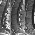
48 Infections MRI is the most sensitive imaging modality for the detection of spondylodiskitis. Figures 48.1A through 48.1C present such a case, with a typical disk space fluid pocket (at L3–L4), seen as high SI on (A) FSE T2WI. Adjacent vertebral bodies are completely involved in this severe infection, but edema in milder cases may be limited to a fraction of the vertebrae adjacent to the disk space–an appearance which, in the absence of disk SI changes or irregularity, appears similar to type 1 degenerative end plate changes. The (A) T2WI and (C
![]()
Stay updated, free articles. Join our Telegram channel

Full access? Get Clinical Tree


