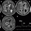
Fig. 15.1
Pneumonia with slow resolution and bilateral infiltrates in a hospitalized and cardiopathic old woman, 12 weeks from clinical onset
15.2.2 Tuberculosis
TB forms slowly, is regarded as chronic, and reactivation of disease, often poorly distinguishable from fibrotic outcomes, is very frequent. In such cases, the symptoms may be nonspecific and not severe, and TB can be confused with a cardiorespiratory disease, chronic bronchitis, or with those diseases generically called “old age” (Pedicelli et al. 2006). Besides, the anatomo-pathological findings are often not pathognomonic of TB, because there can be atypical localizations of infiltrative lesions in the basal areas, infiltrative outbreaks of the primary infection, and nodal involvement, resulting in diagnostic delay and an adverse course.
Chronic pulmonary TB is characterized by alternating episodes of recurrence and remission with endogenous reinfection and spread via the bronchial tree, leading to a real bronchial ubiquitous TB (Leung 1999), characterized by a typical high-resolution CT (HRCT) pattern. The most common complications are bronchiectasis (Fig. 15.2) and the residual cavities in the upper lobes, which in turn can be colonized by the fungus Aspergillus, with formation of typical “fungus ball” or aspergillosis consisting of a coil of hyphae with mucus and cellular debris (Pérez-Guzman et al. 2000).
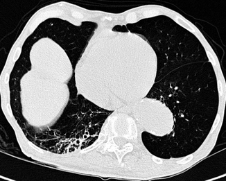

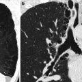
Fig. 15.2
Elderly patient with right lower lobe bronchiectasis, with pulmonary emphysema (see the left lung base), and carrying cutaneous TB with reactivation of a lung infiltrate
The clinical expression characteristic of aspergilloma is hemoptysis. Other complications are many: empyema, due to bronchopleural fistula, rarely documented, and typical of the classic TB, but also present in debilitated subjects, in specific forms, or in chronic nonspecific antibiotic-resistant infections; the revival of an old silent focus in the context of a fibrothorax (Fig. 15.3); pseudoaneurysms of the pulmonary artery, due to erosion from an adjacent tuberculous cavity, which can cause severe hemoptysis; and the occurrence of “scar cancer,” which, although not a proper complication, is worth highlighting as it is more common than cancers arising in a healthy lung and also because it may cause a delay in diagnosis caused by the preexisting fibrotic lesions (Pedicelli et al. 2006).
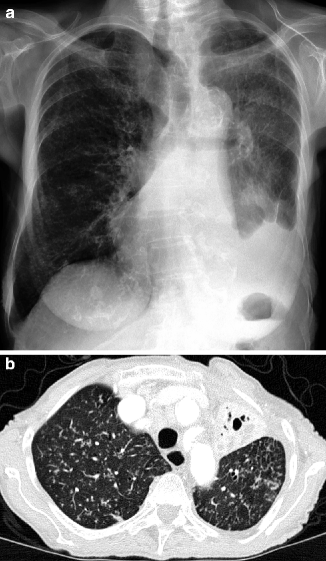

Fig. 15.3
A 89-year-old woman with reactivation of a left apical tuberculosis infiltrate in the context of fibrothorax. (a) CXR. (b) contrast-enhanced CT using a window for parenchyma
Different considerations are necessary with regard to the relation between TB and the pathogen and the consequent therapeutic implications. In the elderly, almost all cases of TB are still due to drug-sensitive strains. It is also important to note that the already treated elderly display a degree of resistance to therapy three times higher than others (Djureti et al. 2002) and there may be a risk of a health emergency due to an increased incidence of multidrug-resistant TB. This has led to a change in the treatment guidelines: in these cases, use of “second-line” antituberculous drugs and of a multidrug therapy – at least three drugs – is recommended (Djureti et al. 2002; Hiyama et al. 2000). Keep in mind the disadvantages of the treatment: longer duration, higher cost, and more toxicity especially in the elderly (McNeil et al. 2003). The effectiveness of an antituberculosis treatment should be evaluated by sputum examination and on monitoring of clinical symptoms and radiographic lesions (Kim et al. 2004). Thus, the absence of clinical and radiological response and presence of positive sputum is regarded as treatment failure, bearing in mind that assessing drug-resistant TB can take up to 6 weeks (Ollè Goig and Sandy 2003). The failure may also be due to noncompliance, intentional or unintentional, by elderly patients or medication error by the medical staff that believes the condition has improved and suspends the treatment too soon (Ollè Goig and Sandy 2003). It should be noted that inadequate or a poorly controlled treatment is more dangerous than no therapy, as it facilitates development of drug resistance.
15.3 Role of Diagnostic Imaging
Some general background information on the role of imaging is necessary. Imaging is called upon to attempt to clarify diagnostic problems, such as differentiating between infectious and noninfectious diseases. Imaging techniques can hardly provide an accurate diagnosis, because the same pathogen can cause different radiological findings and, vice versa, similar radiological findings might result from different conditions. More likely, these methods will show the presence of a disease process and help to narrow the field of diagnostic hypotheses, especially if the radiological presentation is integrated with anamnestic, clinical, and laboratory data (Kim et al. 2004).
Having established these points, imaging does play many roles: demonstrating the presence of a disease process, either clinically suspected or in a routine radiological monitoring assessing morphology, location, and extent; determining whether the condition is infectious or not, and, if the former, proposing a diagnostic orientation or a restricted range of assumptions (bearing in mind also that bronchological techniques may produce nonspecific, if not inaccurate, results in 10–50 % of cases) (Loeb 2003); demonstrating superimposed complications; helping to choose the invasive technique and the most appropriate site for biopsy and bronchoalveolar lavage; and, finally, evaluating the effectiveness of therapy by means of seriated controls.
The diagnostic imaging techniques used in these cases are mainly chest x-rays (CXR) and CT.
As with clinical-epidemiological data acquisition, CXR is the first investigation and is often enough able to orient the diagnosis. Indeed, although radiological negativity with positive clinical objectivity is possible as it is possible to confound pathological processes with the physiological thoracic changes in aging (Schiavon et al. 1993), the sensitivity of CXR is well above that of clinical examination. In its turn, CT – also and especially as HRCT – has a much higher sensitivity and higher specificity than CXR, allowing the following: identification of the pathologic process, especially in cases of discordance between clinical and radiological features that more accurately characterizes its nature; assessment of the true extent of lesions and the presence of complications; and guidance for any invasive procedures (Fein and Niederman 1994).
In TB, CXR is generally not enough to clarify the degree of disease activity, whereas HRCT allows assessment of possible bronchogenic and/or hematogenic spread, of possible extension through the bronchial tree, and of possible persistence or increase of active cavities, highly indicative of multidrug-resistant TB (Fig. 15.4).
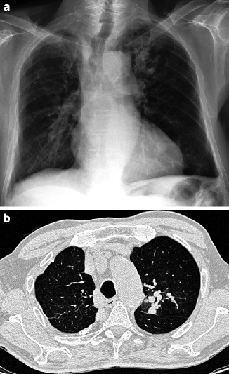

Fig. 15.4
A 77-year-old man with a history of TB admitted with fever and dyspnea. (a) CXR. Alterations, mainly fibrosclerotic, in the left apex. It is not possible to form an opinion on the possible degree of disease activity. In (b), the CT scan shows the chronic fibrosclerotic changes with associated ground glass aspects suggestive of resumption of the disease
15.4 Radiological Findings
15.4.1 Pulmonary Infections
Although radiological findings in most cases are rather nonspecific, there is usually a correlation between the type of radiological pattern and lung disease. Several factors influence the radiological and anatomo-pathological findings of the infection: the routes of access of the germ, its pathogenicity, the mode of intrapulmonary spread of the process, the host’s immune defenses, the coexistence of other diseases, and any established therapy (Wesley Ely and Haponik 1991).
A first distinction is pathological and takes into account the pulmonary structures involved at the onset, the routes of spread, and the type of infiltration (Cametti et al. 1995). Lobar pneumonia is characterized by uniform consolidation crossed by “air bronchogram” for the spread between adjacent alveoli (typical of Streptococcus pneumoniae). Lobular pneumonia or bronchopneumonia is characterized by irregular patchy or nodular opacities, confluent for the spread from the airways to the surrounding parenchyma (typical of Staphylococcus aureus). Interstitial pneumonia is characterized by initial involvement of the peribronchovascular interstitium and by a later spread to the alveoli seen as patchy poorly defined opacities (typical of Mycoplasma pneumoniae and viruses). Hematogenous embolization is characterized by multiple, nodular, and often excavated infiltrates – true pulmonary infarcts – and supported by various germs.
This classification does not always correspond to reality, because the interstitium and the alveolus are part of the same functional unit and it is difficult to consider them separately; at least in the early stages, however, the disease may affect mainly the interstitium or the alveoli, thus facilitating the etiologic diagnosis (Cametti et al. 1995). Therefore, a purely radiological classification based on the exclusive or predominant pattern is more useful and practical: focal, diffuse, or nodular infiltrates.
15.4.1.1 Focal Infiltrates
These are single or multiple opacities, which in the elderly in about 50 % of cases are due to bacterial infections (Streptococcus, gram (−), and Legionella pneumophila) and less commonly to viruses, fungi, and protozoa (Loeb 2004; Canini et al. 1999). The radiological pattern is the classic one of the alveolar syndrome that is poorly defined and confluent acinar opacities with segmental or lobar distribution, with “air bronchogram,” and no apparent collapse (Fig. 15.5). This sign must always be looked for, because it is highly suggestive of lung disease without an obstructive cause – in particular bronchogenic cancer – even if it does not exclude the presence of a neoplasia with lepidic growth like bronchoalveolar carcinoma, which causes great problems of differential diagnosis. The lack of “air bronchogram” on the other side should immediately raise the suspicion of post-obstructive pneumonia by bronchogenic carcinoma (Fig. 15.6).
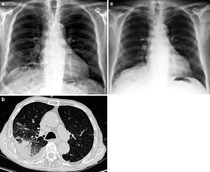
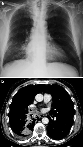

Fig. 15.5
Air bronchogram visible in both CXR and CT in an 80-year-old patient with streptococcal pneumonia (a, b). In (c), the CXR demonstrates resolution of the disease

Fig. 15.6
Post-obstructive consolidation in the superior segment of the right lower lobe, supported by hilar bronchogenic carcinoma in an 80-year-old man. CXR (a) and contrast-enhanced CT (b)
The interlobar fissures may remain in place, be retracted in case of atelectasis, or protrude (“bulging fissure”) from the presence of abundant exudate, as it is a typical appearance in case of Klebsiella or pneumococcal infection (Canini et al. 1999). Complications consist of parenchymal excavation, pleural effusion, and empyema. Excavation prevails in infections caused by Staphylococci and gram-negative bacteria – like the Pseudomonas aeruginosa type (Fig. 15.7


Stay updated, free articles. Join our Telegram channel

Full access? Get Clinical Tree


