22 Inferior Mesenteric Artery
K.I. Ringe, S. Meyer
The inferior mesenteric artery is an artery with a generally constant origin. It nearly always originates from the aorta, usually at the height of the third lumbar vertebra, supplying parts of the transverse colon, the descending colon, sigmoid, and parts of the rectum.1–17 The number of branches to the sigmoid varies considerably from one to six. For the sake of clarity, only two sigmoid arteries are outlined in the respective drawings. The most important end branch of the inferior mesenteric artery is the superior rectal artery, which in 80% of all cases divides into two main branches. These anastomose with the four other rectal arteries, which are branches of the internal iliac artery.3,18 The superior rectal artery is the main arterial blood supply to the rectum. Therefore, a ligature distal to the last anastomosis with a sigmoid artery (the point of Sudeck) can be dangerous. These rectal anastomoses can be of clinical importance when the external iliac artery is occluded and the collateral blood supply of the leg uses the inferior mesenteric, superior rectal, internal iliac, or femoral arteries. In such cases, a mesenteric steal syndrome can be observed.15,19
It is difficult to define an exact border between the areas supplied by the superior and inferior mesenteric arteries because of the arterial arches, the marginal arteries along the colon. In anatomical studies (arterial dissections), the border is normally delimited more orally than in angiographic studies. Angiography shows the functional situation better but gives evidence only for the area supplied by the dye-injected artery and does not exclude an additional supply by another artery. Fig. 22.1 represents a compromise between anatomical and angiographic data.11,16,17,19–25

Fig. 22.1 Schematic demonstration of observed localizations of the watershed area between the superior and the inferior mesenteric artery; the percentages indicate the frequency of each localization.
22.1 The Inferior Mesenteric Artery Branches into the Left Colic Artery, Sigmoid Arteries, and the Superior Rectal Artery (89%)

Fig. 22.2 Trifurcation of the main stem of the inferior mesenteric artery (~25%). Schematic (a) and DSA (b). Besides, urethral contrast excretion can be appreciated (*). 1 Inferior mesenteric artery; 2 left colic artery; 3 sigmoid arteries; 4 superior rectal artery.

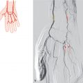
Fig. 22.3 Bifurcation of the main stem of the inferior mesenteric artery; both stems give off branches to the sigmoid (~30%). Schematic (a) and DSA (b). 1 Inferior mesenteric artery; 2 left colic artery; 3 sigmoid branches; 4 superior rectal artery.

Fig. 22.4 Bifurcation of the main stem of the inferior mesenteric artery; the sigmoid arteries originate only from the superior rectal artery (~25%). Schematic.
Related posts:


Stay updated, free articles. Join our Telegram channel

Full access? Get Clinical Tree


