Inferior Vena Cava and Its Main Tributaries

CHAPTER 110 Inferior Vena Cava and Its Main Tributaries


INTRODUCTION


The inferior vena cava (IVC) and major tributary veins are retroperitoneal structures with unique anatomic and developmental characteristics that offer special challenges for clinical and radiologic assessment. Even though the clinical assessment of IVC pathology presents several limitations, the revolutionary advances we have seen in computed tomography (CT) and magnetic resonance imaging (MRI) technology allow us to achieve excellent noninvasive assessments of these structures. The emergence of CT and MRI for vascular imaging has facilitated the transitioning of x-ray catheter angiography from merely a diagnostic tool to a viable less invasive percutaneous therapeutic replacement for complex open surgical interventions.


Multidetector row CT (i.e., MDCT) has become the modality of choice for IVC assessment. The fast scanning speeds that they can obtain has reduced motion artifacts to a minimum and enabled quick extended coverages of body anatomy, notably for rapid assessment of the heart, IVC, and pelvic veins. Another advantage of modern advanced MDCT scanners is their isotropic voxel resolution, allowing improved multiplanar reformation of image data (i.e., axial, coronal, sagittal, or oblique) with high spatial resolution, providing excellent anatomic assessment of complex anatomic relationships that can often be the case when evaluating vascular anatomy of abdominal organs.


MRI assessment of the IVC has also been improved with recent advances. The new phased array coils built with 12 and 16 channels can deliver better coverage of the abdomen and pelvis and provide increased signal-to-noise ratio. It is always important to keep in mind that MRI examinations do not expose the patient to ionizing radiation.


In this chapter, we will discuss the anatomy and pathology of the IVC, starting with the anatomic variants, then we will review tumoral disease affecting the IVC and finally, we will discuss some liver transplantation and interventions.


Inferior Vena Cava


Normal Anatomy and Congenital Anomalies


The IVC extends from the confluence of the common iliac veins at the level of L5 vertebral body, to the right atrium of the heart in right prevertebral location, next to the abdominal aorta and is surrounded by a rich network of lymphatic vessels (Fig. 110-1). It is partially covered anteriorly by the peritoneal membrane. The retroperitoneal space where the IVC is located can communicate with the perirenal spaces and the anterior and posterior interfascial spaces.1
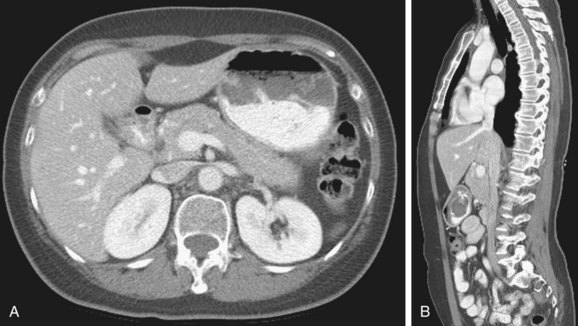


The shape of the IVC varies from round to ovoid or even flat depending on a multitude of factors such as intrathoracic pressure, blood volume status, or the presence of congestive heart failure. The IVC receives a number of tributaries including common iliac, lumbar, renal, right adrenal, and hepatic veins. The IVC lies between the liver and the diaphragm and cephalad courses medially to enter the right atrium. At this level, a fat pad (continuous with the retroperitoneal fat) can be seen in many normal patients in an inferomedial location, sometimes bulging into the lumen of the IVC. This fat should not be considered pathologic and should not generate any further work-up studies.


Congenital anomalies of the IVC generally include abnormal position of the IVC or absence of IVC. The most common IVC anomalies are: (1) left IVC, (2) duplicated IVC, (3) azygos continuation of IVC, (4) circumaortic left renal vein, (5) retroaortic left renal vein, (6) circumcaval or retrocaval ureter, (7) duplicated right renal vein, (8) absence of infrarenal or entire IVC, (9) duplicated IVC with retroaortic right renal vein and hemiazygos continuation of the IVC, and (10) duplication of IVC with retroaortic left renal vein and azygos continuation of the IVC.


Left IVC: The infrarenal IVC is located to the left of the abdominal aorta, then it joins the left renal vein which then crosses anterior to the abdominal aorta and along with the right renal vein forms the normal right-sided prerenal IVC.


Duplicated IVC: There are two IVCs below the level of the renal veins—each connected to the ipsilateral common iliac vein. The left IVC joins the left renal vein, which then crosses anterior to the abdominal aorta and drains into the right IVC (Fig. 110-2). There may be variants in this anatomy and there may be significant discrepancy in the size of the two IVCs.
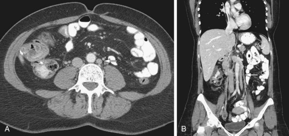


Azygos continuation of IVC: The infrarenal portion of IVC receives blood from the renal veins. It passes posterior to the diaphragmatic crura, enters the thorax as the azygos vein, and then joins the superior vena cava at the azygos arch. The hepatic and right adrenal veins drain directly into the right atrium. The gonadal veins drain into the ipsilateral renal veins. The right renal artery crosses abnormally anterior to the IVC (Fig. 110-3).
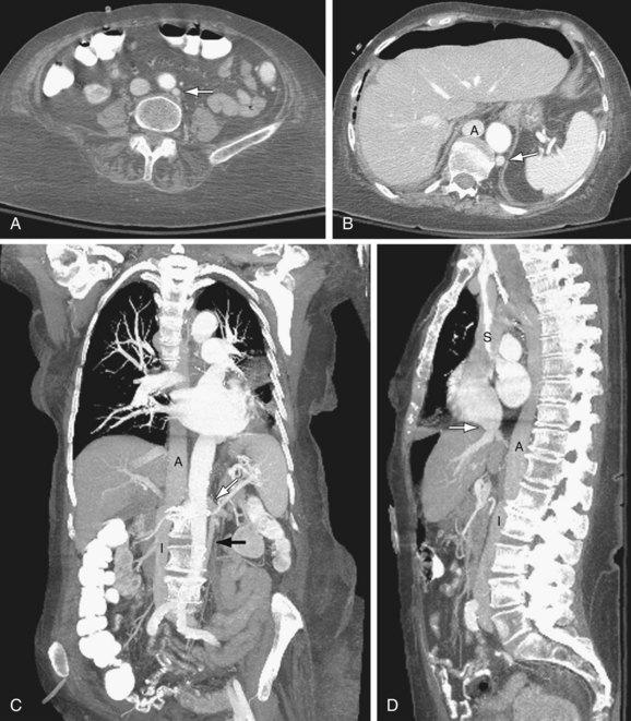


Circumaortic left renal vein: There are two left renal veins. The superior-anterior renal vein receives the adrenal vein and crosses the aorta anteriorly to join the IVC. The second renal vein is approximately 1 to 2 cm more inferior and posterior. It receives the left gonadal vein and crosses posterior to the aorta to join the IVC (Fig. 110-4).
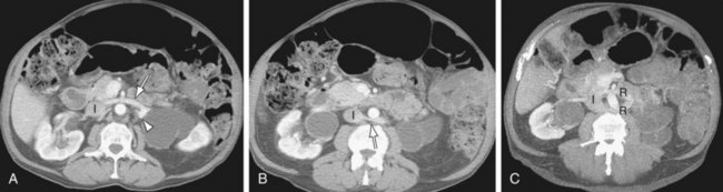


Retroaortic left renal vein: The renal vein crosses posterior to the aorta to join the IVC.


Circumcaval ureter (also known as retrocaval ureter): The anomaly always occurs on the right side. The proximal right ureter courses posterior to the IVC, emerges to the right of the aorta, and lies anterior to the right iliac vessel.


Duplicated right renal vein: There is presence of two right renal veins, one anterior and one posterior, usually at the same level.


Partial or complete absence of IVC: The variants of this anomaly include complete absence of the entire IVC which may include the iliac veins as well and partial absence of IVC with preservation of the suprarenal segment. In either case, the iliac veins join to form enlarged ascending lumbar veins. If the entire IVC is absent, the anterior paravertebral collateral vessels convey the blood return to the azygos and hemiazygos veins. If the suprarenal IVC is present, it receives blood from the renal veins. With partial or complete absence of the IVC, large gonadal and parauterine veins can be seen.


Duplication of IVC with retroaortic right renal vein and hemiazygos continuation of the IVC: There are two IVCs below the level of the renal veins. The right IVC joins the right renal vein, which crosses posterior to the aorta to drain in the left IVC. The left IVC then passes posterior to the diaphragmatic crura and continues into the thorax as the hemiazygos vein. There may be a significant size difference between the two vessels. In the thorax, the hemiazygos vein may have any of these different drainage pathways: (1) it crosses posterior to the aorta at about T8 to T9 to join the rudimentary azygos vein; (2) it joins a persistent left SVC and drains into the coronary vein; (3) an accessory hemiazygos continues to join the left brachiocephalic vein.


Duplication of IVC with retroaortic left renal vein and azygos continuation of IVC: There are two infrarenal IVCs. The left IVC joins the left renal vein which then crosses posterior to the aorta to join the right IVC. It passes posterior to the diaphragmatic crura and enters the thorax as azygos vein. It then joins the superior vena cava at its normal location in the right paratracheal space. The hepatic segment may not be truly absent. It drains directly into the right atrium.


Etiology and Pathophysiology (Including any Special Anatomic Considerations)


Genetic factors may play a role in IVC anomalies because having a first-degree relative with an IVC anatomic anomaly is considered a risk factor.


Embryogenesis


Knowledge of the IVC embryogenesis is necessary for a better understanding of the IVC anatomic aberrations. The IVC is composed of four segments which form during the 6 to 8 weeks postconception.2,3 This is due to continuous appearance and regression of three paired embryonic veins, which include the posterior cardinal veins, the subcardinal veins, and the supracardinal veins. The first step in this complex process is the formation of the posterior supracardinal and more anterior subcardinal veins. Then, the most caudal segment of the right supracardinal vein becomes the infrarenal vena cava. The hepatic segment of IVC is derived from the vitelline vein, which conveys blood from the viscera. The suprarenal segment is formed via a subcardinal-hepatic anastomosis. The renal segment forms via anastomosis of the right supra-subcardinal and post-subcardinal veins. The infrarenal segment arises from the right supracardinal vein.


Congenital anomalies of IVC are due to interruption of normal regression or lack of development of the different segments. The circumaortic venous ring and retroaortic left renal vein are related to aberrant development of the renal segment. Azygos continuation of the IVC results when there is a developmental anomaly involving the suprarenal segment.


Early in embryogenesis, there are two renal veins for each kidney: ventral and dorsal. Normally, the dorsal renal vein involutes as the anterior persists as the main renal vein in adult patients. Anatomic anomalies can occur if the involution of the dorsal veins does not occur, including retrocaval and circumaortic left renal vein and duplication of right renal vein.


Imaging Indications and Algorithm


The symptomatic patients would require evaluation of the venous system in the lower extremity and the urinary system. The best imaging consideration would be CT with multiplanar reformation. The detection of anatomic variants in the renal veins is particularly important at the time of surgical planning for kidney donation. Although the diagnosis of left renal vein variants is easy to detect, in the right kidney the findings of a double vein can be more subtle and sometimes may be overviewed.


Although the diagnosis of IVC anatomic aberrations may be suspected with abdominal ultrasonography, the assessment is usually limited due to their deep location, difficult insonation angle for Doppler studies, and/or the presence of bowel gas that may obscure key segments of the veins. The compression performed during a standard abdominal ultrasonographic examination may also cause collapse of some veins, making the anatomic assessment even more limited. The superior anatomic assessment provided by MRI or MDCT of the abdomen and pelvis makes them the modalities of choice at the time of making the final diagnosis. The advantages of these two modalities are among the following:


There are some risks involved in the use of MDCT, including the ones related to ionizing radiation and the use of intravenous contrast media. MRI risks are related to those associated with the magnetic field but also to that of intravenous contrast agent administration if performed. X-ray catheter angiography is indicated primarily for therapeutic purposes such as installing an IVC filter, taking a biopsy of an intraluminal mass, or installing a stent to treat a venous stenosis.


Imaging Techniques and Findings


TUMORS OF THE IVC AND MAIN TRIBUTARIES


Prevalence and Epidemiology


Primary IVC tumors (leiomyosarcomas) are very rare with only one large series published in the literature.5 It is more common to see tumors invading the IVC through its tributary veins arising from separate abdominal organs. One of the most common causes of neoplastic invasion of the IVC lumen is the renal cell carcinoma (RCC) that can be seen invading the IVC through the renal vein in 4% to 10% of the cases. Hepatocellular carcinoma (HCC) frequently invades the portal vein but also on rare occasions can invade the IVC through the hepatic veins. Primary adrenocortical carcinoma is a rare adrenal tumor that invades the IVC. Multiple other retroperitoneal tumors can compress and invade the IVC, including lymphomas, metastasis of gonadal or uterine tumors, pheochromocytomas, and other retroperitoneal sarcomas.


Dec 26, 2015 | Posted by in CARDIOVASCULAR IMAGING | Comments Off on Inferior Vena Cava and Its Main Tributaries

Full access? Get Clinical Tree

Get Clinical Tree app for offline access