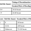
Marc Sapoval, Massimiliano di Primio, Olivier Pellerin, Thierry Carrères and Pierre-Yves Laffy In most countries, critical limb ischemia (CLI) has an incidence estimated to be 50 to 100 per 100,000 every year. CLI has a high mortality and morbidity rate and consumes important health and social care resources.1,2 Aging of the population, increasing incidence of diabetes and renal failure, and limitations in the ability to reduce tobacco consumption explain the high likelihood of increased incidence in the near future. Treatment options include amputation, surgical or endovascular revascularization, and novel techniques of arteriogenesis or angiogenesis using stem cell or gene transfer. Amputation should be avoided as much as possible because it carries its own risk of complications and increased mortality. Bypass surgery requires good-quality saphenous veins (not always present) and routine ultrasound-guided graft surveillance.3 The optimal approach should be selected according to multidisciplinary consensus and is generally based on local and general clinical factors, high-quality anatomic workup, and experience. As stated in the TransAtlantic Inter-Society Consensus (TASC) document in 20001 and confirmed by the recent results of the BASIL trial,4 the endovascular approach is generally regarded as the initial treatment of choice. Based on current evidence, the indication for below-the-knee (BTK) angioplasty is limited to patients presenting with chronic CLI as defined in the TASC consensus document.1 Indications for BTK angioplasty are based on clinical assessment of the patient and evaluation of the extent of arterial disease. Revascularization is the only method that will prevent amputation and prolong life. These patients are at high risk for limb loss and other vascular events like stroke and myocardial infarction (MI). Studies have shown that 25% of patients who cannot be revascularized will die within 1 year after the initial diagnosis of CLI.5,6 MRA has several advantages that are well recognized, but it is limited by the possibility of claustrophobia and other well-known contraindications. MRA for the demonstration of BTK lesions has a sensitivity of about 93%, and with recent technical refinements (venous occlusion cuff, parallel imaging), the results should further improve.7,8 Multidetector CTA (MDCTA) is now rivaling digital subtraction angiography (DSA) in the quality of imaging sensitivity and specificity.9 MDCTA requires iodinated contrast medium, but careful hydration can overcome most of the problems related to nephrotoxicity. A significant limitation is analysis of small and calcified vessels. The results of high-quality MRA and CTA for the diagnosis of BTK disease are broadly comparable. The gold standard for imaging BTK circulation is DSA, which has become easier to perform thanks to the development of smaller-caliber diagnostic catheters and less toxic iodinated contrast media. A unifemoral approach, where only the ipsilateral femoral artery is punctured and subsequent angiography limited to the symptomatic leg, requires substantially less contrast medium than required for classic bilateral angiography. Patients taking metformin or other biguanides should undergo interruption of these medications for at least 48 hours after DSA, with reintroduction only after verification of unimpaired creatinine clearance (European Society of Urological Radiology guidelines).10 In practice, puncture of the ipsilateral common femoral artery (CFA) with a Teflon needle and injection of less than 60 mL of a contrast agent can provide adequate images of the lower limb arterial supply. Delayed images with selective injections, appropriate pixel shifting, and masking are also key elements to allow high-quality images. Equipment needed for endovascular revascularization includes: a. To assist in the crossover technique: pigtail or Cobra, sometimes internal mammary catheter. b. To assist in catheterizing the origin of the superficial femoral artery (SFA): any catheter with an angulated tip (e.g., Bolia Minicath [Terumo Medical Corp., Somerset, N.J.], Vanshi 2 or 3 [Cook Medical, Bloomington, Ind.] Van Andel catheter for subintimal use [Cook Medical]). a. Antegrade approach: 4F or 6F (Terumo): 10, 25, or 40 cm long. b. Crossover: first choice is KSAW-5.0 (Cook Medical) with a removable Y-connector that can be replaced with a hemostatic valve. The Shuttle-SL flexor sheath (up to 80 cm) (Cook Medical) is also useful, as is the Destination sheath 5F to 6F (Terumo). a. Intraluminal recanalization: first choice is the Pilot 200 (Abbott Vascular, Santa Clara, Calif.). Other dedicated 0.014-inch wires such as the guide approach CTO (chronic total occlusion) (Cook Medical), Winn (Abbott Vascular), and V14 (Boston Scientific, Natick, Mass.). Coronary CTO 0.014-inch wires such as PT Graphix (Boston Scientific) and Choice PT (Boston Scientific) can be helpful in selected cases. b. Subintimal recanalization: hydrophilic 0.035-inch J and straight (stiff in some cases) (Terumo) and previously mentioned 0.014-inch wires advanced in a loop shape. c. 0.014-inch wires for calcified occlusions (Cross-it [Abbott Laboratories, Abbott Park, Ill.]; Pilot intermediate [Guidant, Santa Ana, Calif.]). 5. Contrast media (low-osmolar contrast media preferred because of frequent renal failure in these patients): Xenetix 320 (Roissy, France) or Visipaque 320 (GE Healthcare Inc, Princeton, N.J.). a. Size 0.014- or 0.018-inch coaxial balloons are preferable because they allow better crossability of calcified lesions (lower profile). Small (1.25 or 2 mm) balloons can be used to predilate difficult lesions. Short coronary balloons have better lesion crossability (Ryujin [Terumo]). b. Size 0.035-inch balloons are preferred in subintimal angioplasty because they provide better pushability and allow progressive dilation by very short iterative inflation/deflation. c. High-pressure (up to 30 atm) balloons (Dorado [Bard Peripheral Vascular, Tempe, Ariz.]) are useful to overcome recoil in subintimal recanalization. d. 0.035-inch ReKross Catheter (Bard PV) is useful in a very tight subintimal space. a. Self-expandable dedicated stents: Xpert (Abbott Vascular) self-expandable thin-strut nitinol stent, Maris Deep (Medtronic Inc., Minneapolis, Minn.) thin-strut nitinol stent, Astron Pulsar (Biotronik, Bulack, Switzerland). b. Coronary balloon expandable stents: PRO-Kinetic explorer (Biotronik, Berlin, Germany), cobalt chromium (CoCr) and passive coating (silicon carbide), diameter 2.0-5.0 mm, length 8-40 mm. c. Dedicated balloon-expandable stents: Chromis Deep (Medtronic Inc.) CoCr stent up to 8 cm in length, Inperia (Maquet, Orleans, France) (Not available in the U.S.).
Infrapopliteal Revascularization
Indications
Equipment
Imaging
Endovascular Revascularization
Infrapopliteal Revascularization