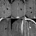
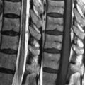
2 Infratentorial Brain Neoplasms Medulloblastoma, primarily a pediatric tumor, is the most common infratentorial neoplasm; 75% originate in the cerebellar vermis (Figs. 2.1A,B). Though medulloblastomas were once thought to be a type of primitive neuroectodermal tumor (PNET), studies have shown their molecular profile to be distinct. Medulloblastomas are highly malignant, commonly spreading via the cerebrospinal fluid (CSF). Thus, lumbar puncture and contrast-enhanced MRI of the entire neural axis are essential with a medulloblastoma. Metastasis outside the CNS is rare, most commonly involving the bone marrow. Treatment consists of some combination of resection, craniospinal radiotherapy, and chemotherapy. Like most brain tumors, medulloblastomas have low SI on T1WI (white arrow, Fig. 2.1A). On T2WI, their appearance varies from isointense to mildly hyperintense relative to brain parenchyma. Medulloblastomas show restricted diffusion, a point of distinction from astrocytomas. Intense, heterogeneous contrast enhancement (Fig. 2.1B, black arrow) is typical, although some lesions demonstrate only patchy enhancement. Medulloblastomas tend to extend into the 4th ventricle, leading to obstructive hydrocephalus, reflected in part by ventricular enlargement in Fig. 2.1A. The increase in ventricular pressure leads to compensation by transventricular absorption, with a thin uniform band of abnormal high SI present on T2WI in the periventricular white matter (Fig. 2.1C, white arrow
![]()
Stay updated, free articles. Join our Telegram channel


Full access? Get Clinical Tree


