CHAPTER 5 Inner Ear
Embryology
The maturation of the inner ear involves three main phases:
1. Development (4th to 8th week)
2. Growth (8th to 16th week)
3. Ossification (16th to 24th week)
The development of the sensory epithelium within the membranous labyrinth occurs simultaneously with growth and ossification (8th to 24th week). The inner ear arises from a plate-like thickening of the surface neuroectoderm located between the first branchial groove and the hindbrain, the “otic placode.” Each otic placode then invaginates and sinks below the surface neuroectoderm into the underlying mesenchyme, resulting in formation of the “otic pit.” The edges of the otic pit fuse to form the otic vesicle (otocyst). The otic vesicle is the precursor of the membranous (or endolymphatic)labyrinth.
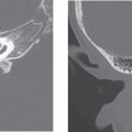
When the embryo is 6 to 7 mm in length, the otic vesicle divides into a larger utricular portion and the smaller endolymphatic portion. The dorsally located utricular portion later forms the utricle, semicircular canals, and endolymphatic duct. The ventral saccular portion differentiates into the saccule and the cochlear duct. The primordial cochlear pouch, which develops as an evagination of the saccule, starts to elongate and begins to coil. By the end of the 8th week, the entire membranous labyrinth is identifiable, and formation of the 2½ to 2¾ cochlear turns is complete. The fetus is able to hear with maturation of the organ of Corti, which occurs by the 24th week of gestation (Fig. 5-1).
The development of membranous labyrinth is completed by the third trimester. Maturation of sensory end organs occurs first in the utricle and saccule, followed by the semicircular canals and, finally, the cochlea. The cochlea is the last part of the membranous labyrinth to undergo maturation and is therefore more subject to developmental malformations compared with vestibular system. Once the membranous labyrinth has matured (6 to 7 months of fetal age), no further growth occurs during the remaining lifetime of the individual. The only exception is the endolymphatic duct and sac, which continue to grow and reach their mature size after puberty.
The otic capsule is the precursor to the bony labyrinth. The otic capsule develops as a cartilaginous condensation of mesenchyme around the otic vesicle between the 4th and 8th weeks of gestation. Growth of the otic capsule continues to the 16th gestational week. During its growth, vacuoles begin to arise in the cartilaginous otic capsule. These vacuoles eventually coalesce to form the perilymphatic space. The newly created perilymphatic space contains perilymph fluid, which surrounds and bathes the membranous labyrinth. Ossification of the otic capsule occurs between weeks 16 and 24 via 14 ossification centers. The end result is the “bony labyrinth.”
Otic capsule ossification is unique in that there are a large number of ossification centers—14—considering the small size of the final product; there is no epiphyseal growth (centers fused directly); and maturation is arrested in a primary state of ossification, that is, endochondral bone persists.
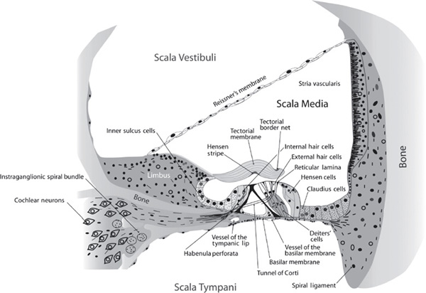
Figure 5-1 Schematic illustration of the anatomy of the cochlea and organ of Corti. (See Color Plate 5-1)
Anatomy of the Inner Ear
The inner ear labyrinth consists of bony and membranous components. The membranous labyrinth contains endolymphatic fluid and it serves as an organ of hearing and balance. The membranous labyrinth is suspended in perilymphatic fluid, which in turn is encased by the bony labyrinth. The bony labyrinth can be divided into three components: the vestibule, the semicircular canals, and the cochlea (Fig. 5-2).
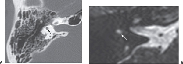
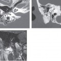
Figure 5-2 (A) Axial computed tomography (CT) and (B) T2-driven (T2 DRIVE) equilibrium pulse sequence image through the superior aspect of the epitympanum demonstrates the crux of the superior semicircular canal (arrows).
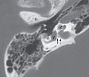
Figure 5-3 Axial CT through the epitympanum obtained inferior to Figure 5-2, demonstrating the vestibule (V), cochlea (C), and canal for the superior vestibular nerve (arrows).
The Vestibule
The vestibule is a central ovoid cavity within the bony labyrinth, located lateral to the fundus of the internal auditory canal. The vestibule plays a vital role in static balance and is innervated by branches from the vestibular nerve along its medial wall and the floor. The vestibule houses the saccular and utricular portions of the membranous labyrinth and displays a high T2 signal within the signal void of the temporal bone on the magnetic resonance (MR) imaging. The vestibule communicates anteriorly with the cochlea and posteriorly with the semicircular canals (Fig. 5-3 and Fig. 5-4).
The Cochlea

Stay updated, free articles. Join our Telegram channel

Full access? Get Clinical Tree


