Instability
Jeffrey S. Ross, MD
Key Facts
Terminology
Loss of spine motion segment stiffness, when applied force produces greater displacement than normal, with pain/deformity
Imaging
Deformity, which increases with motion and over time
Various parameters used to measure degenerative instability by plain films
Dynamic slip > 3 mm in flexion/extension
Static slip of ≥ 4.5 mm
Angulation > 10-15° suggests need for surgical intervention
Flexion-extension plain films best for definition of motion
Top Differential Diagnoses
Pseudoarthrosis
Infection
Endplate destruction, disc T2 hyperintensity
Tumor
Enhancing soft tissue mass
Postoperative
Following multilevel laminectomy or facetectomy
Pathology
Degenerative instabilities
Axial rotational
Translational; plain films show spondylolisthesis, traction spurs, vacuum phenomenon
Retrolisthesis; plain films show increased retrolisthesis with extension
Degenerative scoliosis
Post laminectomy; resection of 50% of bilateral facets alters segmental stiffness
Post fusion; altered biomechanics
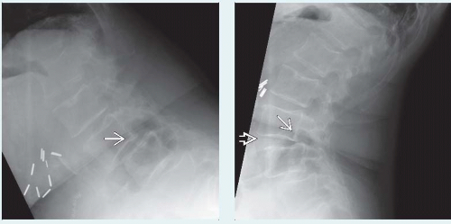 (Left) Wide laminectomy destabilizing spine is shown. Flexion lateral radiograph demonstrates grade 2 anterolisthesis  at L3-4 that improves on extension (see next image). (Right) Lateral radiograph in extension shows grade 2 anterolisthesis, which is mildly reduced at L3-4 that improves on extension (see next image). (Right) Lateral radiograph in extension shows grade 2 anterolisthesis, which is mildly reduced  compared to the flexion deformity. Note the widening of the anterior disc space with extension compared to the flexion deformity. Note the widening of the anterior disc space with extension  . . |
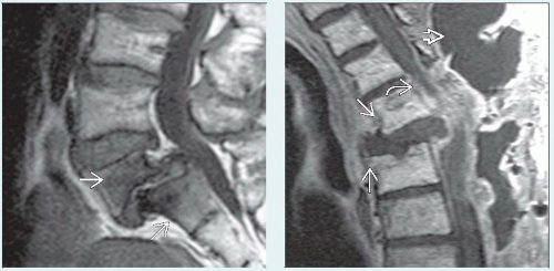 (Left) Sagittal T1WI MR in an adolescent shows severe spondylolisthesis of L5 on S1, with disc degeneration and extensive type I degenerative endplate changes  . (Right) Sagittal T1WI C+ MR shows progressive deformity and retrolisthesis . (Right) Sagittal T1WI C+ MR shows progressive deformity and retrolisthesis  at operative site following extensive posterior debridement for infection, marked thecal sac compression from the deformity and residual phlegmon at operative site following extensive posterior debridement for infection, marked thecal sac compression from the deformity and residual phlegmon  , and extensive dorsal pseudomeningocele , and extensive dorsal pseudomeningocele  . . |
TERMINOLOGY
Synonyms
Spine instability (SI), segmental instability, abnormal spinal motion, degenerative instability
Definitions
Loss of spine motion segment stiffness, when applied force produces greater displacement than normal, with pain/deformity
IMAGING
General Features
Best diagnostic clue
Deformity that increases with motion and over time
Location
Any spinal motion segment (composed of 2 adjacent vertebrae, discs, and connecting spinal ligaments)
Size
Displacement may vary from few mm to width of vertebral body
Morphology
Displacement of vertebral body with respect to adjacent body
Stabilizing anatomic structures
Ligaments
Anterior longitudinal ligament
Resists hyperextension
Posterior longitudinal ligament
Intertransverse ligaments
Connect neighboring transverse processes
Interspinous ligaments
Resist hyperflexion
Facet capsule
Ligamentum flavum
Intervertebral disc
Main stabilizer of lumbar and thoracic spine
Muscular attachments
Both global (rectus and abdominal muscles) and local paraspinal muscle groups
Radiographic Findings
Radiography
Various parameters used to measure degenerative instability by plain films
Dynamic slip > 3 mm in flexion/extension
Static slip ≥ 4.5 mm
Angulation > 10-15° suggests need for surgical intervention
Traction spurs
Vacuum phenomenon
Fluoroscopic Findings
Increased motion with flexion/extension or translation
CT Findings
NECT
Nonspecific findings of degenerative disc disease ± spondylolisthesis
MR Findings
T1WI
Anterolisthesis, retrolisthesis, lateral translation
Nonspecific changes of degenerative disc disease
Controversial as to role in defining instability: Type I degenerative endplate changes
T2WI
Loss of disc signal ± disc space height
STIR
Type I endplate changes may be more evident on this sequence
T1WI C+
Nonspecific enhancement of disc due to degenerative disc disease
Enhancement of type I degenerative endplate changes
Imaging Recommendations
Best imaging tool
Flexion-extension plain films
Protocol advice
MR findings useful as secondary tool for degeneration, endplate changes, stenosis, and herniation
DIFFERENTIAL DIAGNOSIS
Pseudoarthrosis
Abnormal low T1 signal extending through disc, posterior elements, and ligaments
Infection
Endplate destruction, disc T2 hyperintensity
Tumor
Enhancing soft tissue mass
Postoperative
Following multilevel laminectomy or facetectomy
PATHOLOGY
General Features
Etiology
Multifactorial
Associated abnormalities
Clinical and imaging relationship of disc degeneration to instability controversial
Many causes of spinal instability
Fractures
Infection (especially anterior column involvement)
Primary bone and metastatic tumors (vertebral body destruction, neural compression, post resection)
Isthmic spondylolisthesis (L5-S1 progressive deformity in children)
Scoliosis
Degenerative instabilities
Axial rotational (recurrent pain worse with twisting)Related posts:
 Pseudomembranous Colitis (Clostridium Difficile)
 Imaging with VHF Signal Generation: A New Contrast Mechanism for Cancer Imaging Over Large Fields of View
 Pathologic Findings in Aortic Stenosis
 Introduction of Emerging Infectious Diseases
 Regulation of PET Radiopharmaceuticals Production in Europe
 Musculoskeletal Imaging

Stay updated, free articles. Join our Telegram channel

Full access? Get Clinical Tree


