Intermetatarsal Bursitis
Anatomic Considerations
In a manner analogous to that of the digital nerves of the hand, the digital nerves of the foot travel through the intrametatarsal space to innervate each toe. The plantar digital nerves, which are derived from the posterior tibial nerve, provide sensory innervation to the major portion of the plantar surface (Fig. 23.1). These nerves are subject to entrapment and resultant development of perineural fibrosis and degeneration resulting in the clinical syndrome known as Morton neuroma (Fig. 23.2). The dorsal aspect of the foot is innervated by terminal branches of the deep and superficial peroneal nerves. The
overlap of the innervation of these nerves may be considerable. The intermetatarsal bursa lies between the metatarsophalangeal joints in a position that is just dorsal to the interdigital nerves (Fig. 23.3). The bursae extend approximately 1 cm beyond the distal border of the ligament in the web spaces between the second and third and third and fourth digits (Fig. 23.4).
overlap of the innervation of these nerves may be considerable. The intermetatarsal bursa lies between the metatarsophalangeal joints in a position that is just dorsal to the interdigital nerves (Fig. 23.3). The bursae extend approximately 1 cm beyond the distal border of the ligament in the web spaces between the second and third and third and fourth digits (Fig. 23.4).
 FIGURE 23.1 Anatomy of the metatarsophalangeal and digital nerves. |
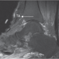
 FIGURE 23.2 Patients suffering from Morton neuroma present with the complaint of pain in the plantar surface with associated dysesthesias radiating into the adjacent toes. |
Clinical Correlates
Intermetatarsal bursitis refers to a constellation of symptoms including pain and tenderness over the affected intermetatarsal spaces which radiate distally into the toes especially if the adjacent interdigital nerve is inflamed. The pain of intermetatarsal bursitis is exacerbated by weight bearing and wearing high heels or shoes which are too narrow. Obesity may also predispose to this condition. The patient suffering from intermetatarsal bursitis is often unable to stand on tiptoes or walk up stairs. Walking and standing for long periods makes the pain worse. The pain of intermetatarsal bursitis is constant and is characterized as sharp and may interfere with sleep. Coexistent neuritis, neuropathy, Morton neuroma formation, stress fractures, metatarsalgia, and synovitis may confuse the clinical picture (Fig. 23.5). As the bursitis worsens, the affected intermetatarsal bursae tend to expand surrounding the adjacent interdigital nerves making the patient’s clinical presentation indistinguishable from the pain of Morton neuroma. If the inflammation of the intermetatarsal bursae becomes chronic, calcification of
the bursae and fibrosis of the surrounding interdigital space may occur.
the bursae and fibrosis of the surrounding interdigital space may occur.
 FIGURE 23.3 Anatomy of the intermetatarsal bursa. |
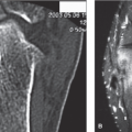
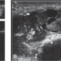
 FIGURE 23.4 Morton neuroma. Axial T1-weighted (A) and postcontrast fat-suppressed T1-weighted (B) images demonstrating neuromas between the second and third and third and fourth metatarsal bases. C: Illustration of the location of Morton neuromas and intermetatarsal bursae with relation to the transverse metatarsal ligament. (From Berquist TH. Imaging of the Foot and Ankle. 3rd ed. Philadelphia, PA: Lippincott Williams & Wilkins; 2011:200.) |
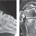
 FIGURE 23.5 A: Radiograph shows subtle cortical thickening to the second metatarsal shaft consistent with a stress fracture. B: Bone scintigram confirms this to be consistent with a fracture. STIR (C) and T1-weighted (D) MR images confirm the associated edema and cortical thickening. (From Pope TL Jr, Harris JH Jr, eds. Harris & Harris’ The Radiology of Emergency Medicine. 5th ed. Philadelphia, PA: Lippincott Williams & Wilkins; 2013:961.) |
 FIGURE 23.6 Mulder maneuver is accomplished by firmly squeezing the two metatarsal heads together with one hand while placing firm pressure on the interdigital space with the other hand. |
On physical examination, pain can be reproduced by squeezing the affected web space between the index finger and thumb. If the interdigital nerve is involved or if a Morton neuroma has developed, a positive Mulder sign can be elicited by firmly squeezing the two metatarsal heads together with one hand while placing firm pressure on the interdigital space with the other hand (Fig. 23.6). The patient with intermetatarsal bursitis often exhibits an antalgic gait in an effort to reduce weight bearing during walking.
Related posts:

Stay updated, free articles. Join our Telegram channel

Full access? Get Clinical Tree


