CHAPTER 36 Internal Auditory Canal Stenosis/Atresia
Epidemiology
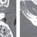
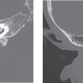
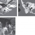
An internal auditory canal (IAC) measuring less than 2 mm in vertical dimension by high-resolution temporal bone computed tomography (CT) is considered stenotic. Approximately 12% of patients with congenital sensorineural hearing loss have radiographic evidence of inner ear abnormalities including IAC stenosis. IAC stenosis without other abnormalities is extremely rare.
Clinical Features
Patients with IAC stenosis may present with congenital sensorineural hearing loss caused by aplasia of the vestibulocochlear nerve during development, but in very rare cases it can be asymptomatic.
Pathology
In addition to primary causes of IAC narrowing, osseous lesions such as an exostosis or an osteoma can cause secondary narrowing of the IAC. Also, underlying skeletal disease causing hypertrophy of the surrounding bone (Paget’s disease or otosclerosis) can contribute to IAC stenosis. Hyperpneumatization of the petrous apex can also result in abnormal narrowed conditions.
Treatment
In most cases of congenital sensorineural hearing loss caused by IAC stenosis, hearing devices or cochlear implantation may not be helpful. In asymptomatic IAC stenosis, palliative IAC decompression of cochlear nerve can be considered if sudden onset of deafness occurs.
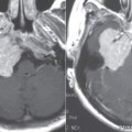
Imaging Findings
CT
Related posts:

Stay updated, free articles. Join our Telegram channel

Full access? Get Clinical Tree


