Numerous systems have been created to divide the mediastinum into specific compartments for the purposes of generating a focused differential diagnosis for masses and other abnormalities identified on imaging, planning for biopsies and surgical interventions, and facilitating communication between health care professionals in a multidisciplinary setting. Most have focused on imaging and are based on arbitrary landmarks delineated on the lateral chest radiograph. The International Thymic Malignancy Interest Group has developed a classification system based on cross-sectional imaging, defining specific prevascular, visceral, and paravertebral compartments, that has been accepted as a new standard and is the topic of this review.
Key points
- •
The classification of mediastinal compartments developed by the International Thymic Malignancy Interest Group (ITMIG) has been designed for use with cross-sectional imaging examinations and is considered a standard.
- •
The ITMIG model divides the mediastinum into 3 unique compartments: the prevascular compartment, visceral compartment, and paravertebral compartment.
- •
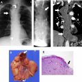
Abnormalities originating from the prevascular compartment tend to be thymic abnormalities, germ cell neoplasms, lymphoma, metastatic disease involving lymph nodes, and goiter.
- •
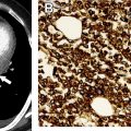
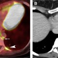
Lesions arising in the visceral compartment include lymphadenopathy due to primary lymphoma or metastatic disease, foregut duplication cysts, tracheal lesions, esophageal neoplasms and vascular lesions originating from the heart, pericardium, and great vessels.
- •
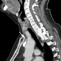
Most abnormalities originating from the paravertebral compartment are neurogenic neoplasms, infections (discitis/osteomyelitis), or those related to trauma.
Introduction
Division of the mediastinum, a region of the thorax that contains many important vascular and nonvascular organs and other anatomic structures, into specific compartments on imaging examinations has been performed for many years by clinical radiologists. Experience has shown that this exercise is extremely valuable when identifying and characterizing masses and other mediastinal abnormalities, and for ultimately determining the next step in clinical management. Several classification schemes have been created not only by radiologists but anatomists and surgeons, although their use has not been uniform. The Shields scheme has been the model most frequently used in clinical practice, whereas the Fraser and Paré’s, Felson, Heitzman, Zylak, and Whitten models have been used by radiologists in clinical practice.
The limitations of existing classification schemes have been well-documented and include significant variations in the terminology and methods used for the identification of individual compartments, which has resulted in miscommunication between health care professionals and the inability to reliably localize some lesions to a specific compartment, and, on the imaging side, the use of arbitrary nonanatomic divisions of the thorax that are based principally on the lateral chest radiograph. In response to the need for a model of the mediastinal compartments designed for use with cross-sectional imaging techniques such as computed tomography (CT), MR imaging, and PET/CT, which are the imaging modalities primarily used in the identification and characterization of mediastinal masses, a new, modern classification scheme has been developed by the International Thymic Malignancy Interest Group (ITMIG), building on prior work by the Japanese Association for Research on the Thymus (JART). In this article, the ITMIG model is illustrated with representative examples.
International thymic malignancy interest group model definitions
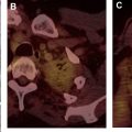
The rationale for and methodology used by ITMIG in the development of this classification scheme has been previously described. , The model developed by ITMIG includes 3 unique compartments: the prevascular compartment (corresponding to the anterior compartment on chest radiography), the visceral compartment (corresponding to the middle compartment on chest radiography), and the paravertebral compartment (corresponding to the posterior compartment on chest radiography) ( Table 1 ). The specific boundaries of each department and the anatomic organs and structures that they normally contain can be easily identified on cross-sectional imaging examinations ( Fig. 1 ). Understanding this model enables the radiologist to predict what lesions are most likely to originate from specific mediastinal compartments.
| Compartment | Boundaries | Contents |
|---|---|---|
| Prevascular | Superior: thoracic inlet Inferior: diaphragm Anterior: sternum Lateral: parietal mediastinal pleura Posterior: anterior aspect of the pericardium as it wraps around the heart in a curvilinear fashion | Thymus Fat Lymph nodes Left brachiocephalic vein |
| Visceral | Superior: thoracic inlet Inferior: diaphragm Anterior: posterior boundaries of the prevascular compartment Posterior: vertical line connecting a point on each thoracic vertebral body 1 cm posterior to its anterior margin | Nonvascular: trachea, carina, esophagus, lymph nodes Vascular: heart, ascending thoracic aorta, aortic arch, descending thoracic aorta, superior vena cava, intrapericardial pulmonary arteries, thoracic duct |
| Paravertebral | Superior: thoracic inlet Inferior: diaphragm Anterior: posterior boundaries of the visceral compartment Posterolateral: vertical line against the posterior margin of the chest wall at the lateral margin of the transverse process of the thoracic spine | Thoracic spine Paravertebral soft tissues |
Related posts:
Stay updated, free articles. Join our Telegram channel

Full access? Get Clinical Tree


